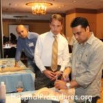
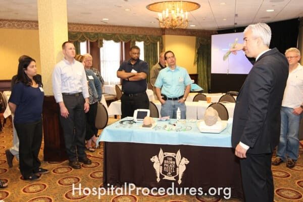
Photos From the Field
click on a title below to see slideshow.


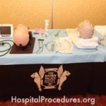


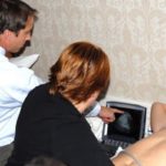
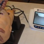
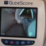
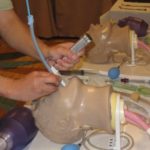
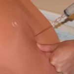
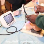
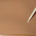
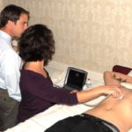
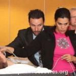
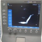
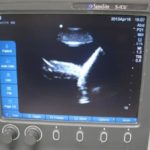
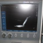
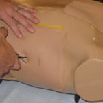
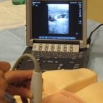
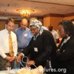
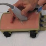
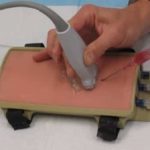
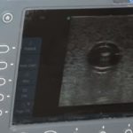
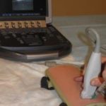
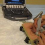
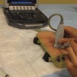
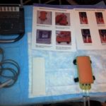
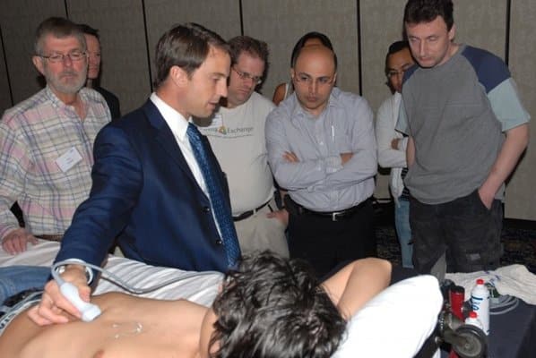
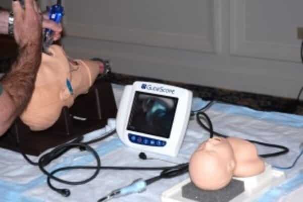
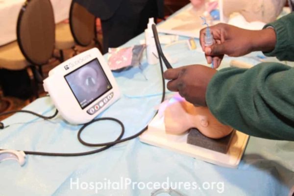
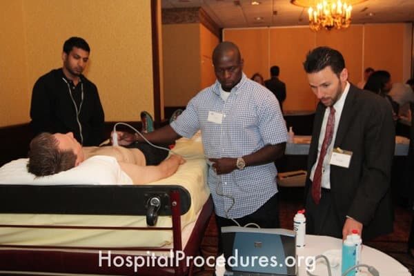
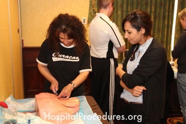
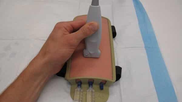
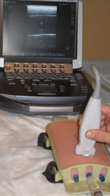
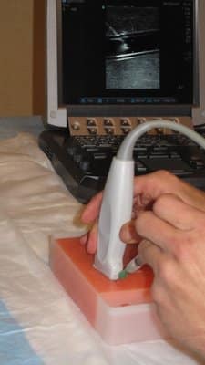
Subxiphoid four chamber cardiac view in the extended-focused abdominal sonography in trauma
E-FAST technique
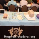
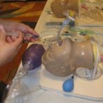
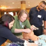
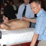
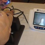
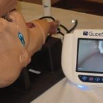
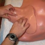
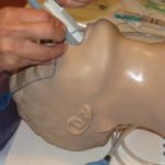
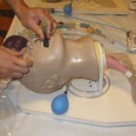
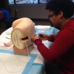
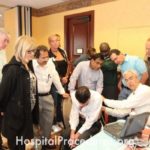
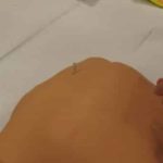
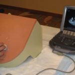
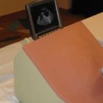
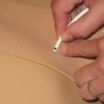
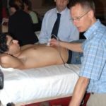
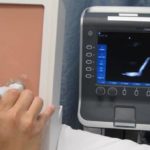
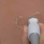
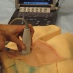
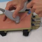
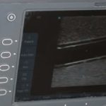
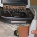
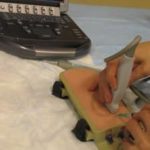
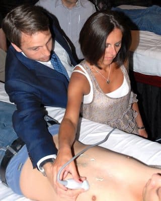
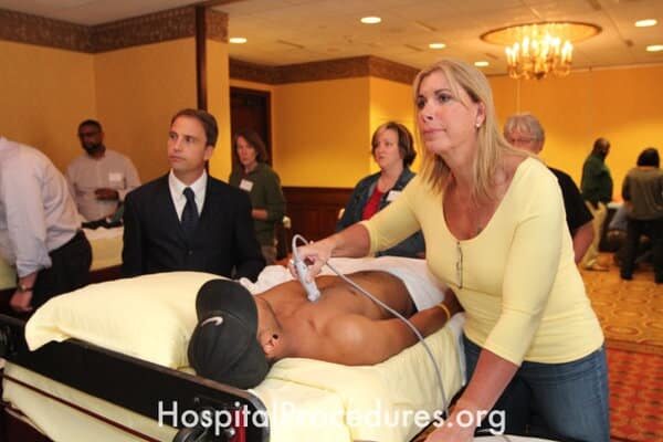
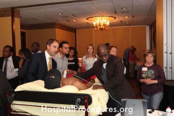
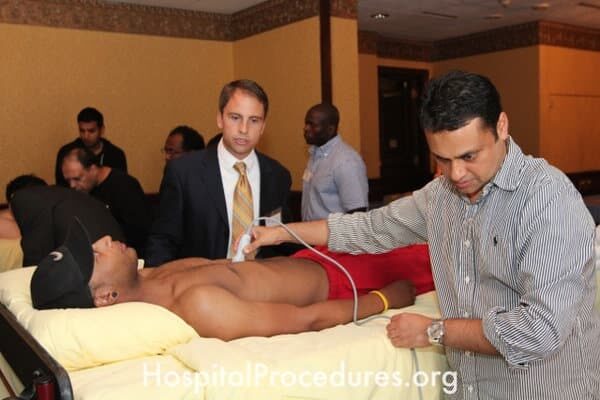
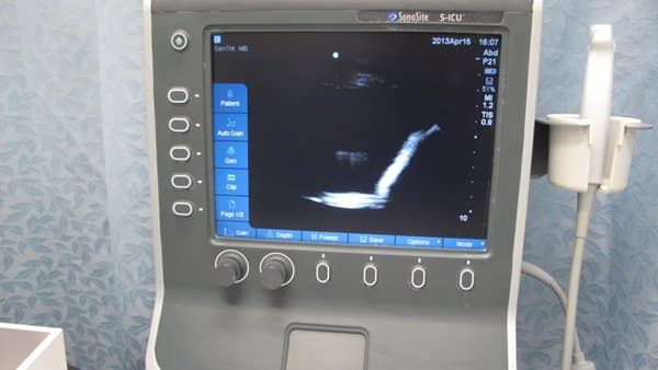
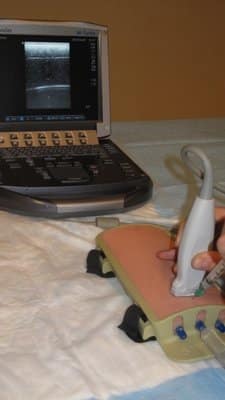
View of the spleen, diaphragm and pleura during teaching of the left upper quadrant E-FAST exam
EFAST LUQ
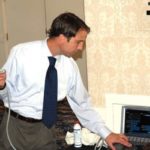
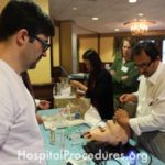
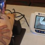
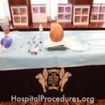
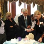
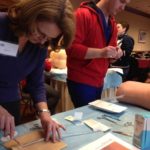
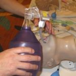
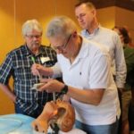
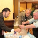
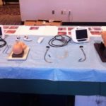
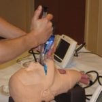
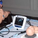
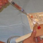
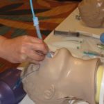
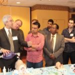
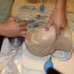
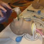
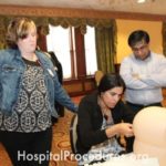
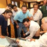
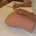
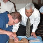
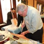
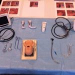
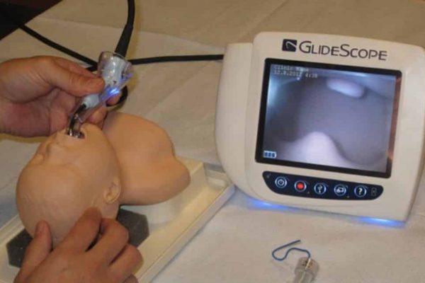
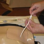
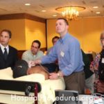
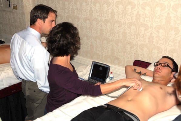
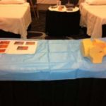
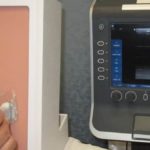
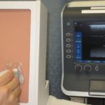
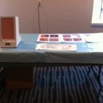
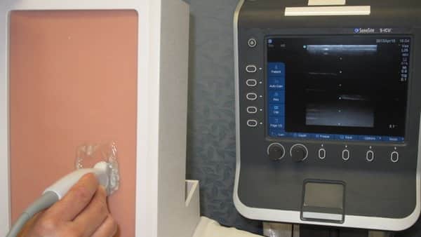
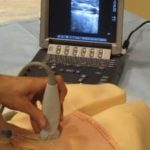
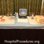
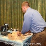
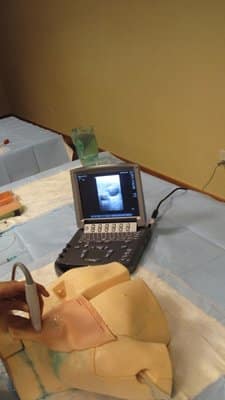
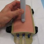
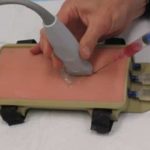
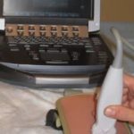
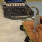
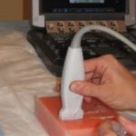
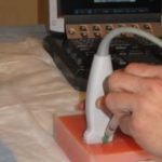
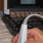
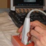
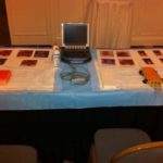
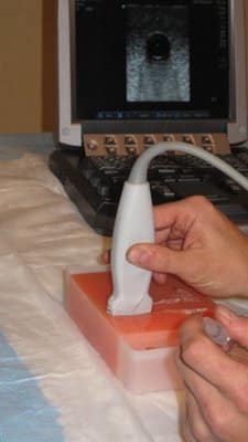
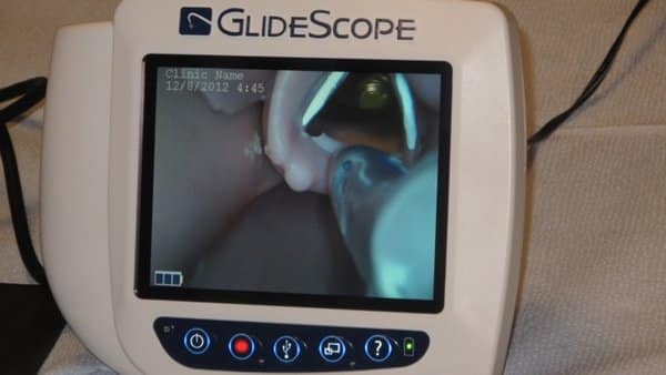
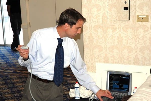
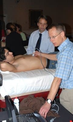
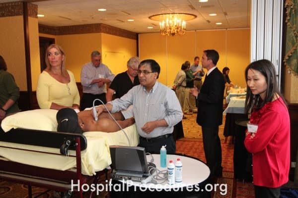
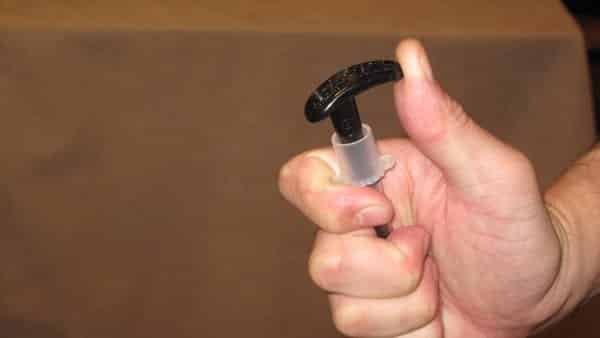
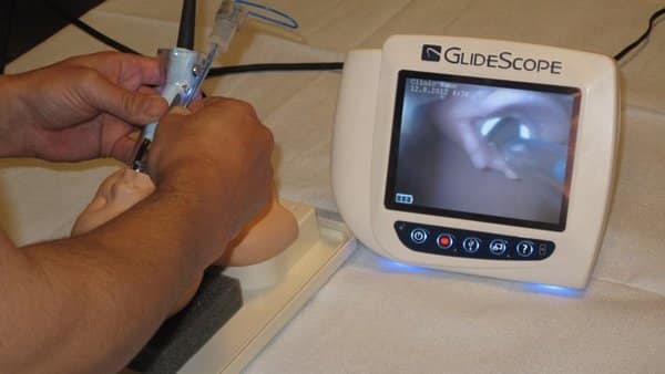
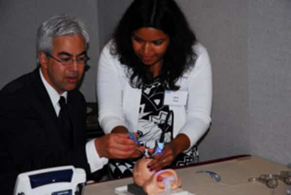
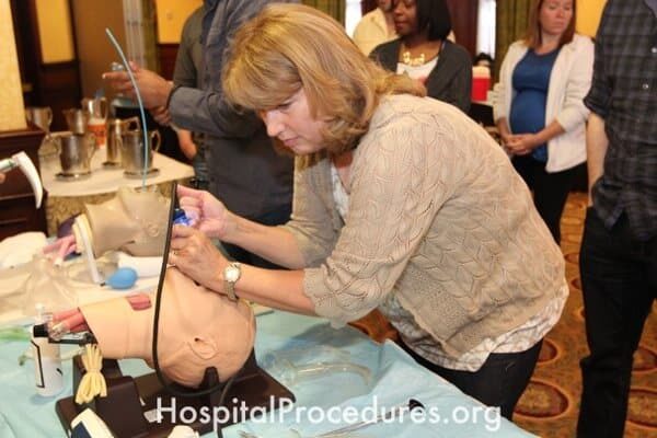
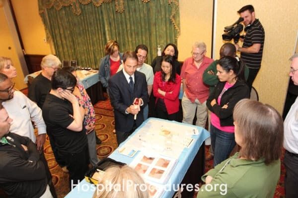
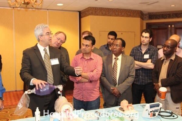
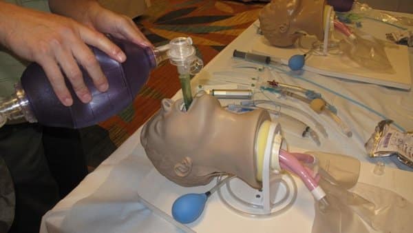
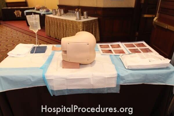
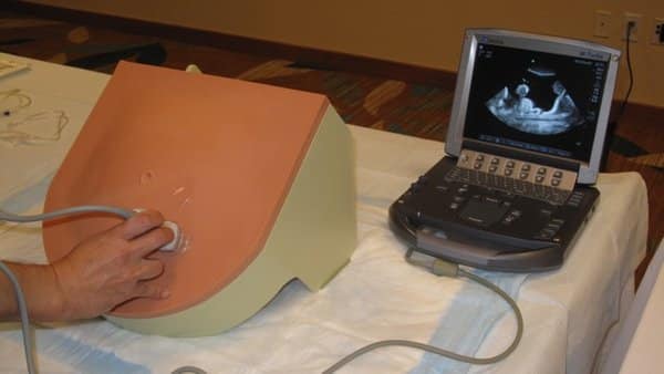
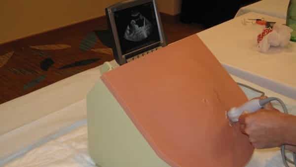
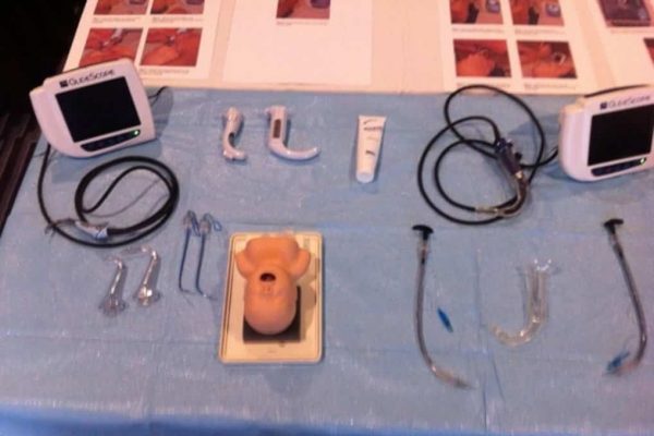
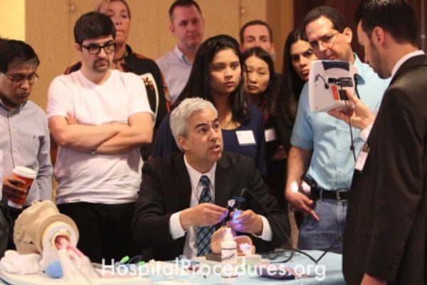
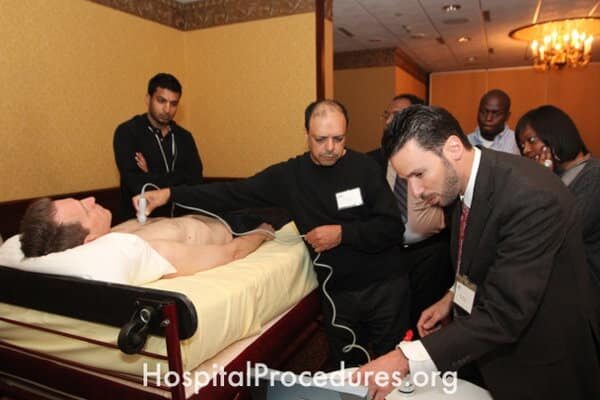
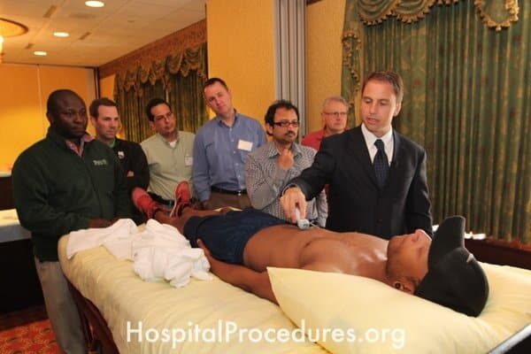
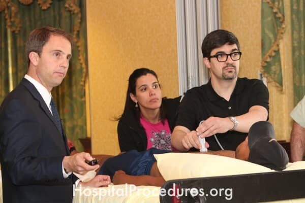
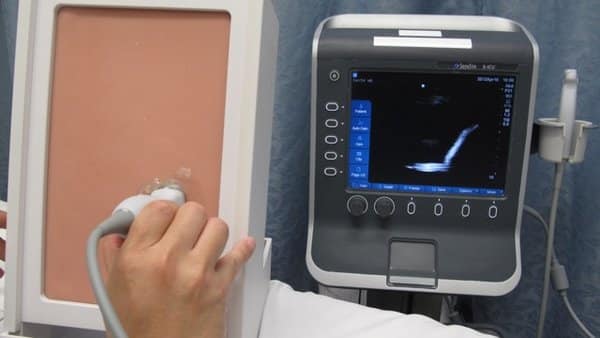
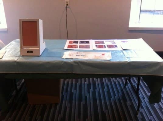
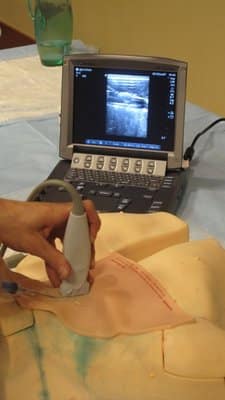
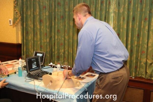
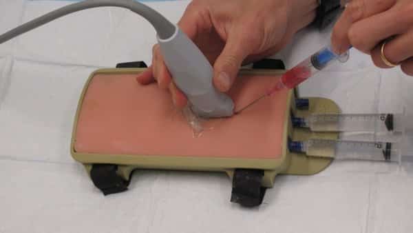
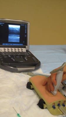
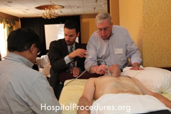
Setting ultrasound depth, gain, probe selection, exam type, probe type and ultrasound mode for E-FAST exam
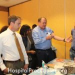
Ultrasound CME

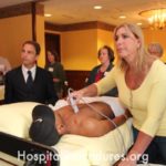


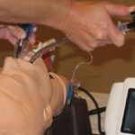
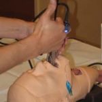

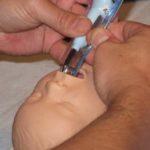


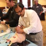


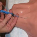
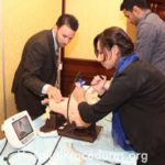
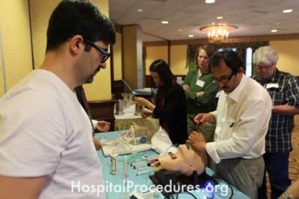
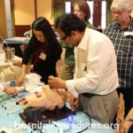
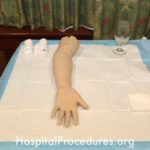
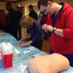
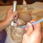
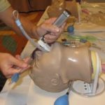
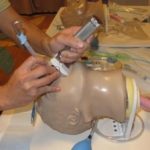
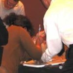
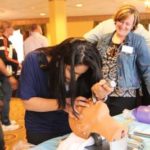
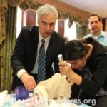
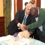
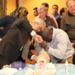
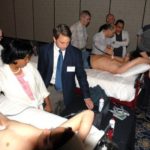
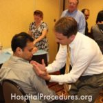
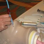
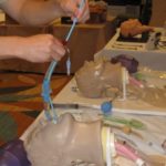
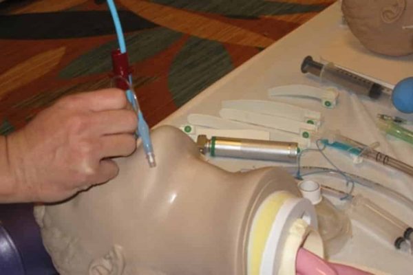
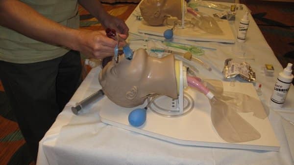
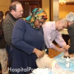
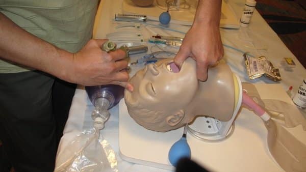
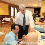
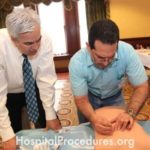
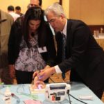
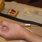
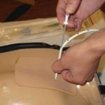
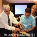
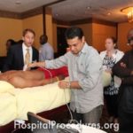
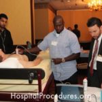
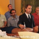
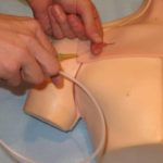
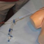
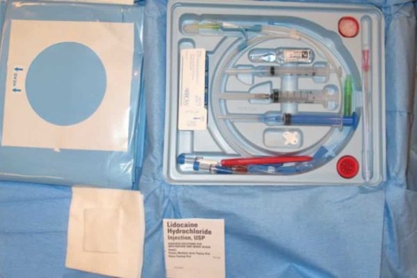
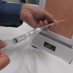
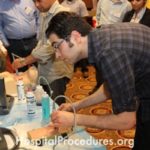
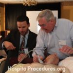
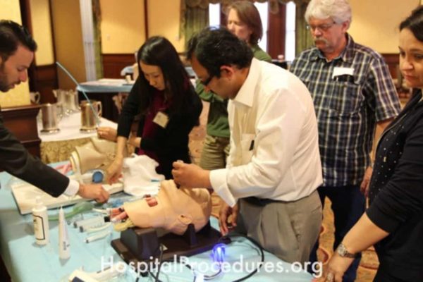
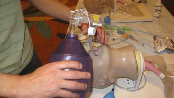
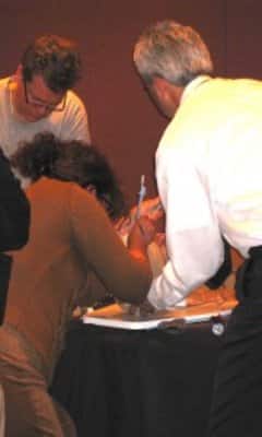
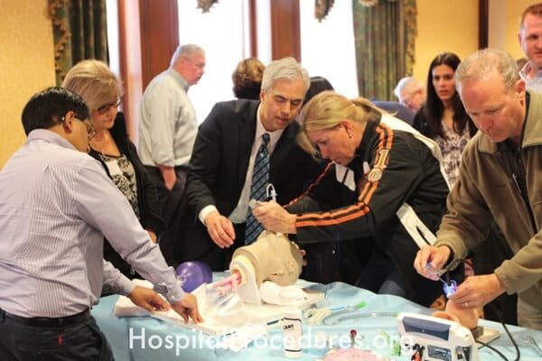
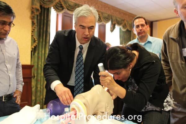
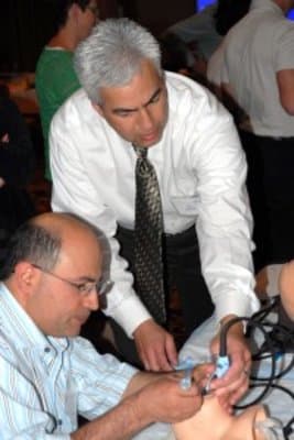
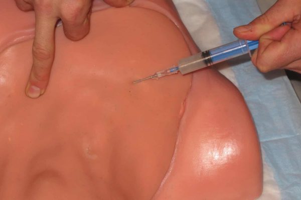
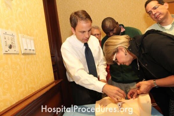
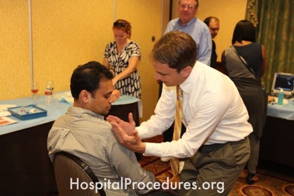
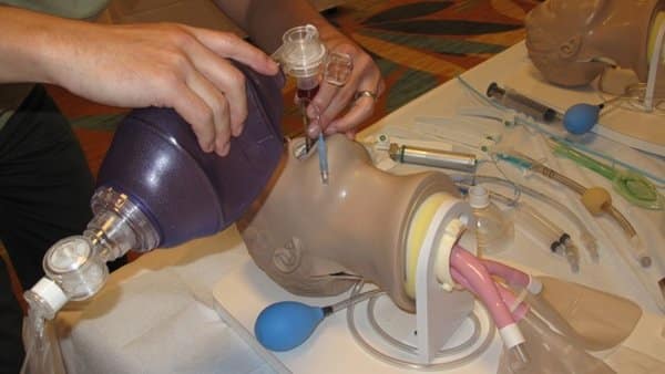
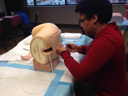
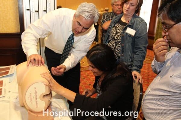
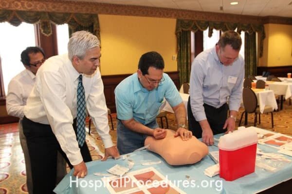
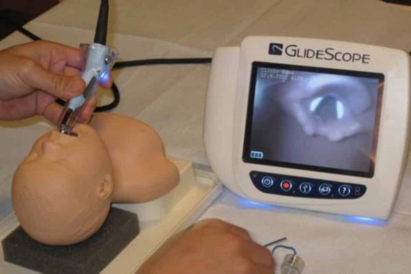
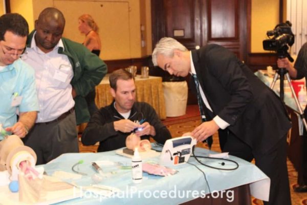
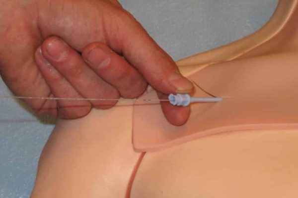
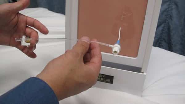
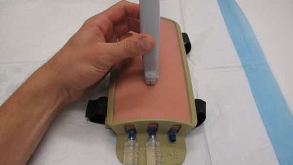
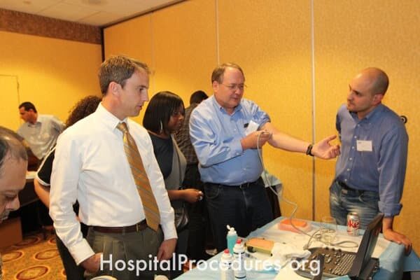
Internal jugular vein central venous catheter placement training
The image captures a training session focused on the placement of a central venous catheter in the internal jugular vein. This essential training addresses the needs of patients requiring hemodynamic monitoring, rapid volume resuscitation, administration of therapies like vasopressors, and vesicant or central parenteral nutrition infusions. The procedure is particularly beneficial for patients with challenging venous access. The training equips healthcare professionals with the skills necessary to accurately and safely insert a central venous catheter in the internal jugular vein, enhancing patient care.
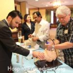
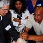
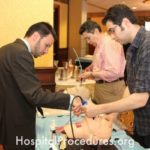
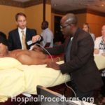
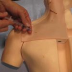
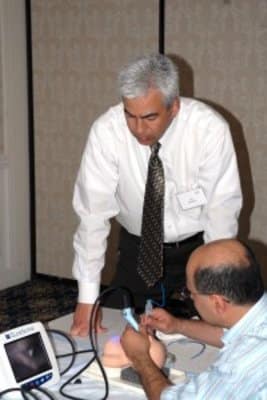
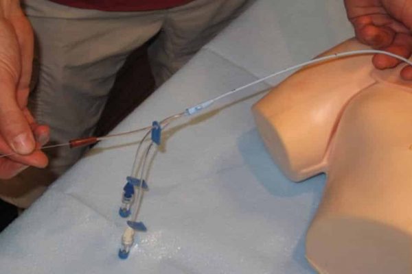
Subclavian central venous catheter placement training
The image captures a training session focused on the placement of subclavian central venous catheters. This training is essential for healthcare professionals to effectively perform central line placement, a procedure necessary for patients requiring hemodynamic monitoring, rapid volume resuscitation, administration of vasopressors, central parenteral nutrition, and vesicant infusions. The training aims to equip medical practitioners with the skills needed to address challenging venous access situations.
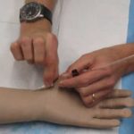
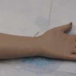
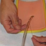
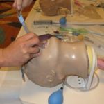
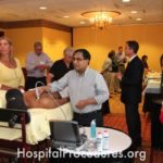
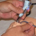
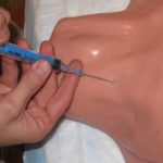
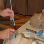
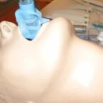
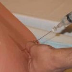
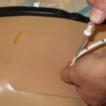
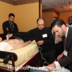
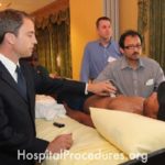
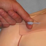
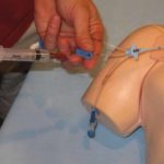
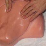
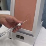
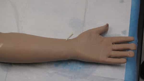
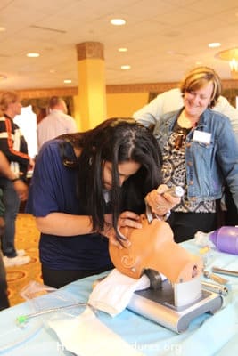
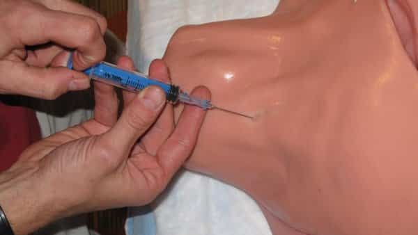
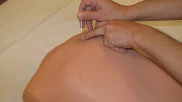
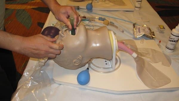
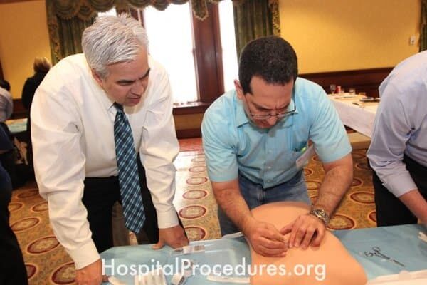
Internal Jugular line
The image depicts the insertion of an internal jugular central line, a procedure crucial for patients requiring hemodynamic monitoring, rapid volume resuscitation, administration of therapies like vasopressors, vesicant or central parenteral nutrition infusions, and for those with challenging venous access. This technique ensures access to a major vein in the neck, enabling effective delivery of essential treatments while addressing critical patient care needs.
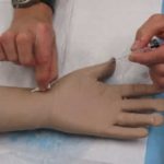
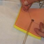
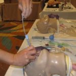
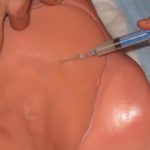
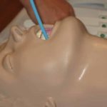
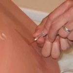
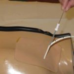
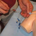
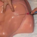
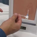
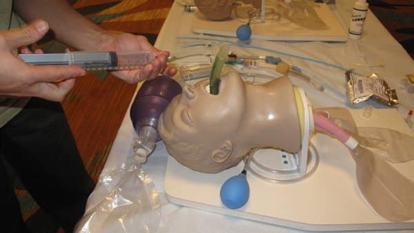
Internal Jugular line insertion
The image illustrates the process of internal jugular line insertion, a critical procedure for patients requiring hemodynamic monitoring, rapid volume resuscitation, administration of therapies like vasopressors, and vesicant or central parenteral nutrition infusions. This technique is especially valuable for patients with challenging venous access. The image captures the essential steps and equipment involved in ensuring successful central line placement in the internal jugular vein.


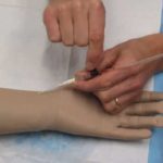
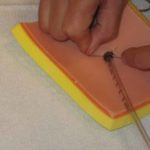
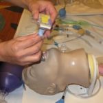
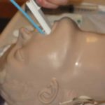
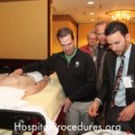
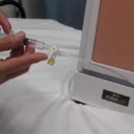
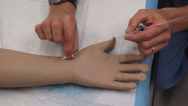
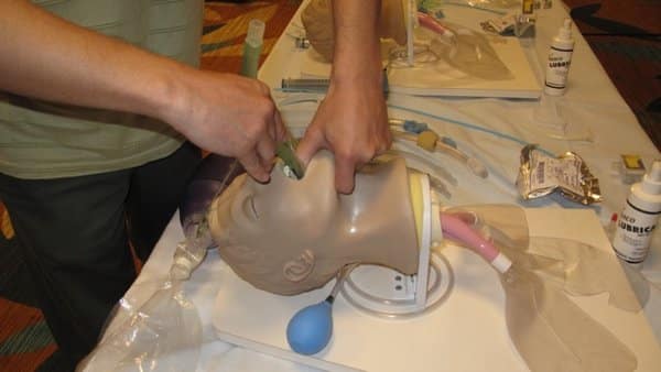
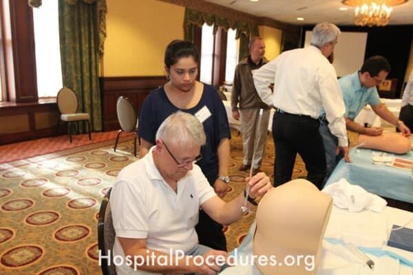
Internal Jugular line placement
The image showcases an Internal Jugular Line Course, designed to educate medical professionals on the placement of internal jugular central lines. This procedure is vital for patients requiring hemodynamic monitoring, rapid volume resuscitation, administration of therapies like vasopressors, and vesicant or central parenteral nutrition infusions. The course addresses the needs of patients with challenging venous access, equipping healthcare providers with the skills to proficiently insert internal jugular central lines, ensuring optimal patient care.
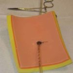
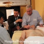
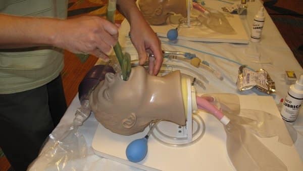
Internal Jugular Line Course
The image portrays an Internal Jugular Line Course, dedicated to educating medical professionals on the technique of internal jugular central line placement. This method is crucial for patients requiring hemodynamic monitoring, swift volume resuscitation, administration of therapies like vasopressors, vesicant or central parenteral nutrition infusions, and for patients with challenging venous access. The course provides comprehensive training, empowering healthcare practitioners to confidently and safely perform internal jugular central line insertions to meet critical patient care needs.
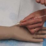
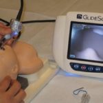
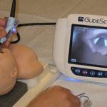
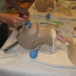
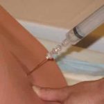
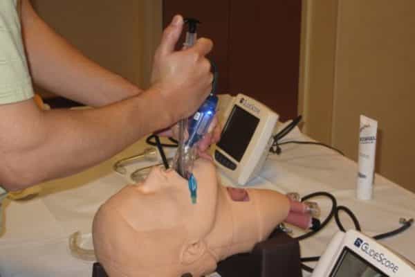
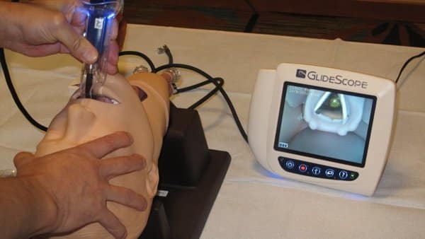
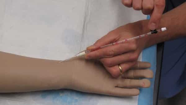
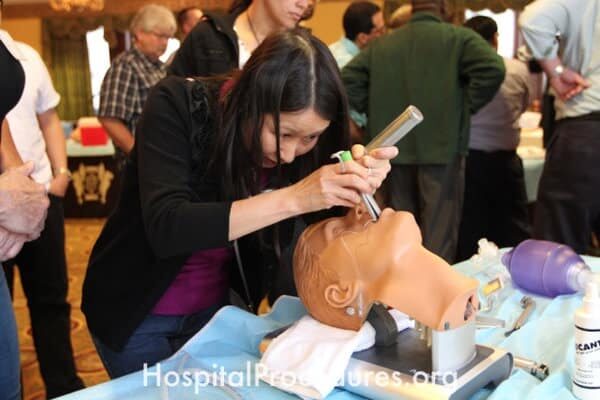
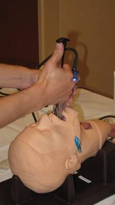
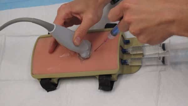
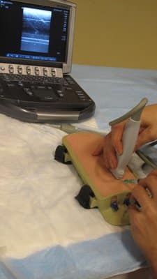
Ultrasound-Guided Internal Jugular Line Placement
The image showcases the technique of ultrasound-guided internal jugular line placement, a critical procedure for patients requiring hemodynamic monitoring, rapid volume resuscitation, administration of therapies like vasopressors, and vesicant or central parenteral nutrition infusions. This method is particularly beneficial for patients with challenging venous access, allowing precise and safe central line insertion into the internal jugular vein. Ultrasound guidance enhances accuracy and ensures optimal patient care.
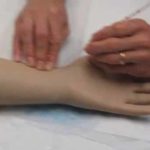
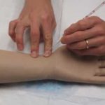
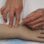
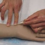
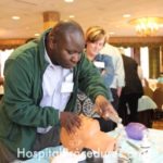
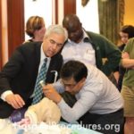
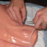
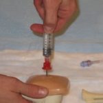
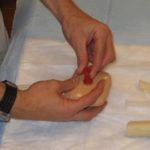
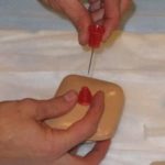
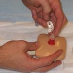
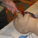
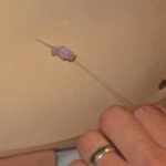
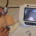
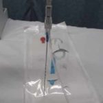
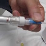
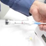
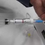
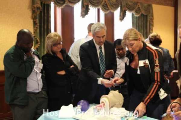
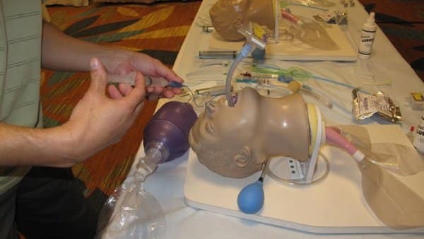
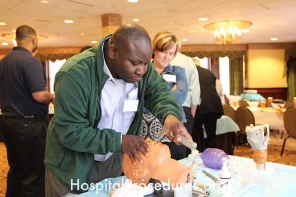
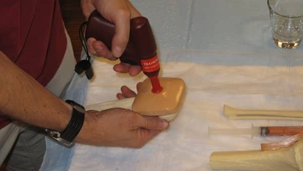
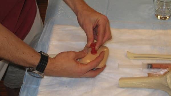
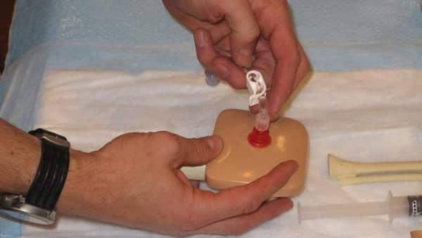
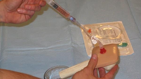
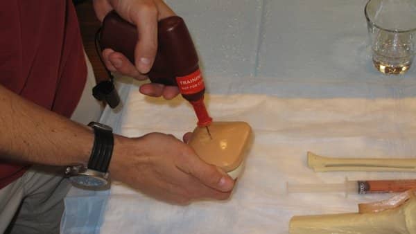
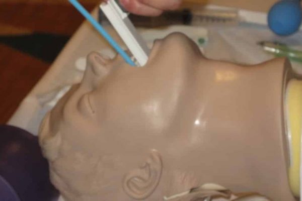
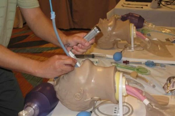
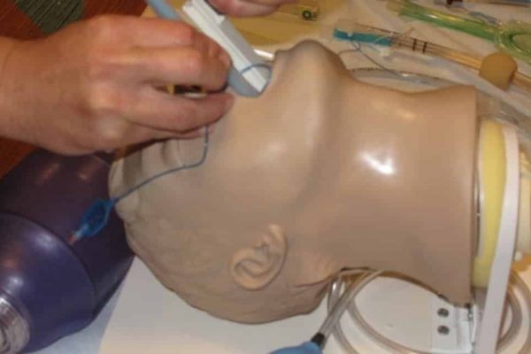
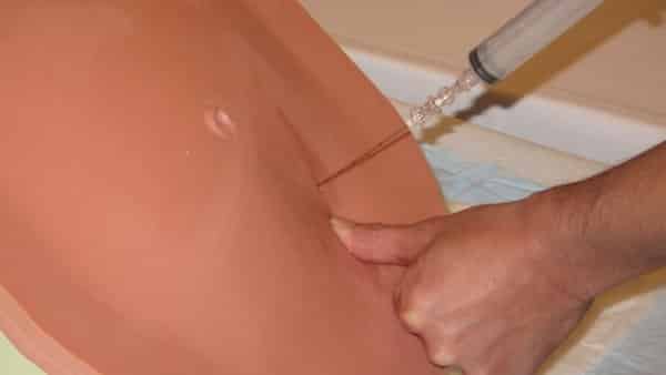
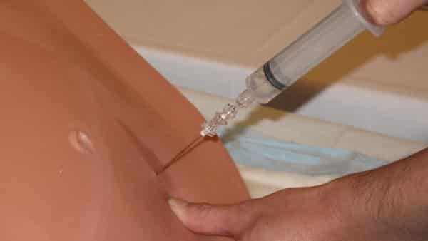
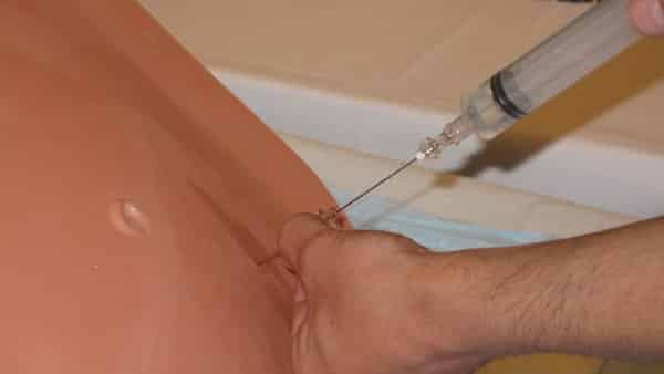
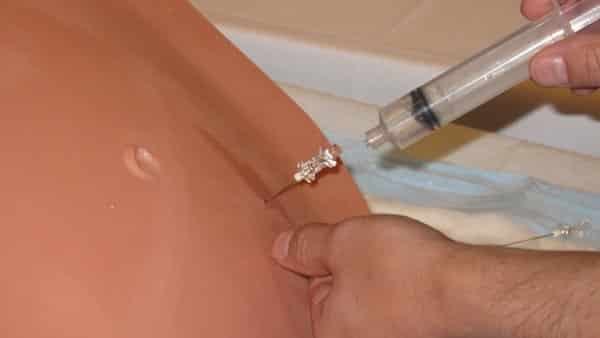
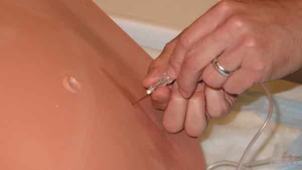
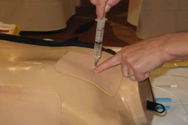
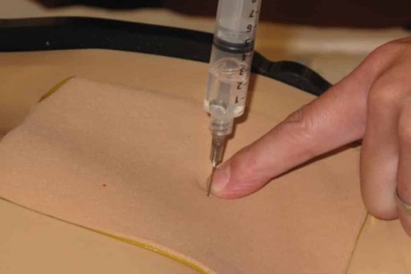
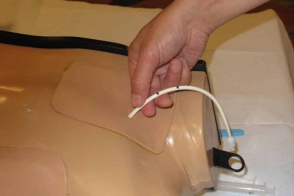
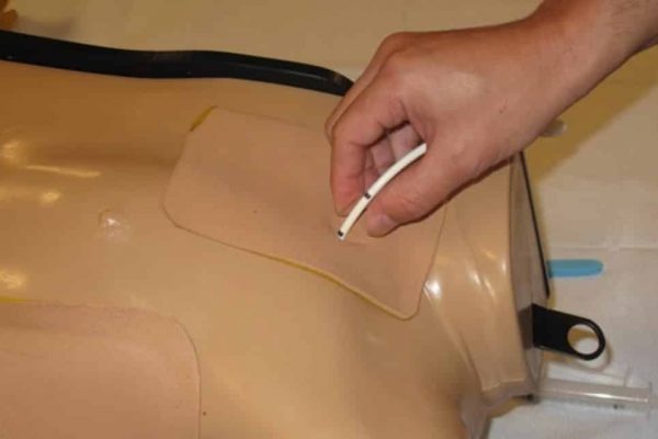
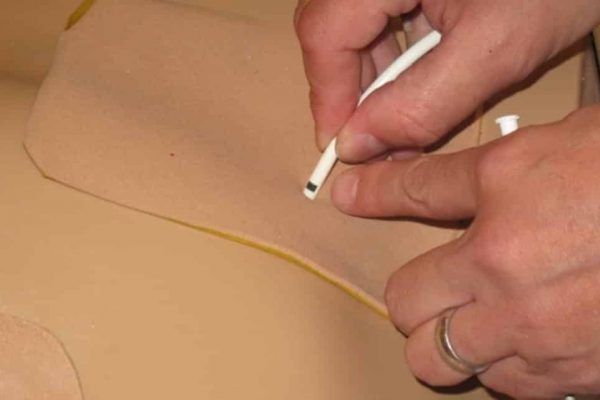
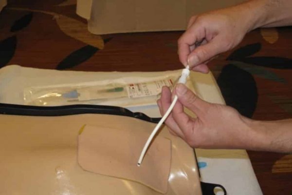
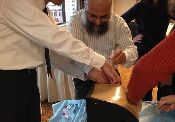
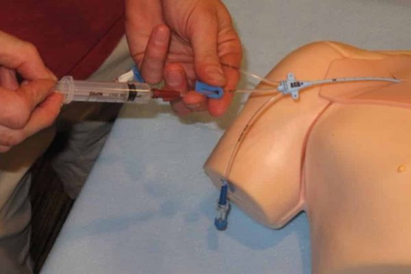
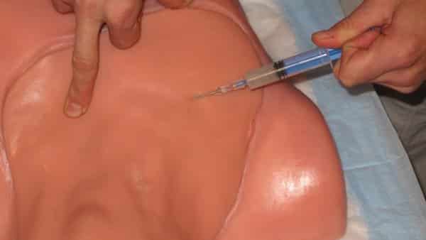
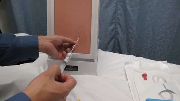
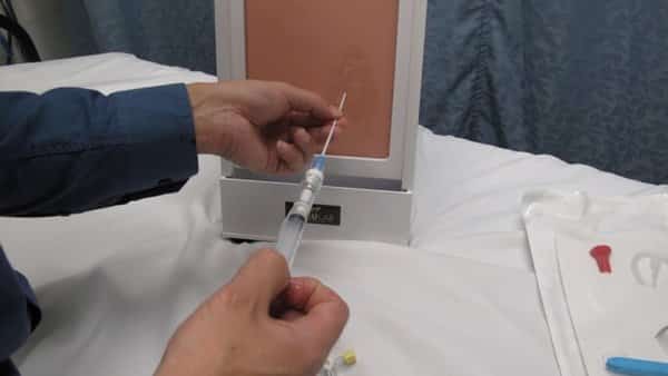
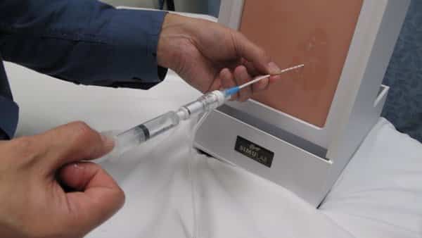
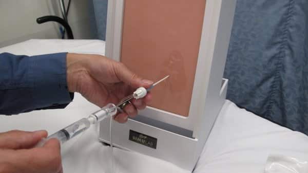
Intraosseous Line Training
The image represents an Intraosseous Line Course, dedicated to teaching the technique of intraosseous line placement. This method serves as an alternative to central venous access, ultrasound-guided central line placement, or ultrasound-guided peripheral IV access, specifically designed for patients facing challenges in vascular access. The course emphasizes the importance of this procedure, providing healthcare professionals with the skills necessary to ensure effective and timely vascular access for patients with difficult venous conditions.
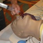
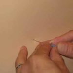

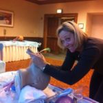
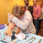
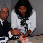
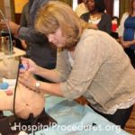
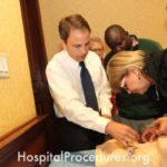
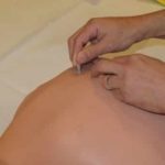
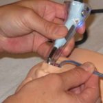
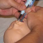
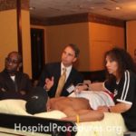
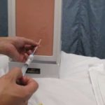
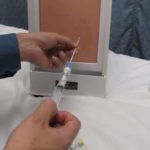
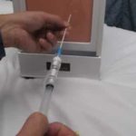
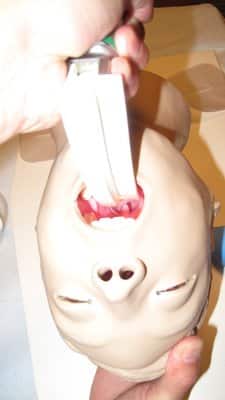
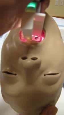
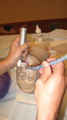
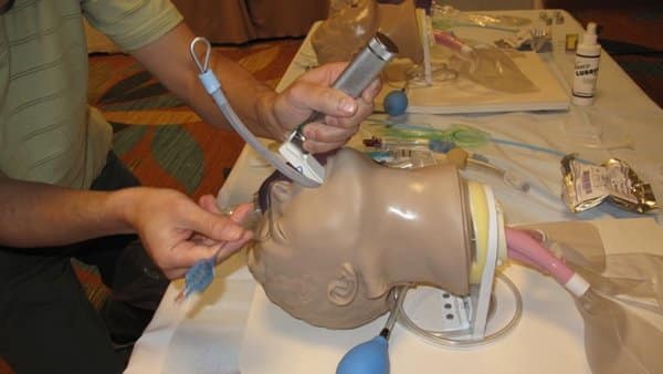
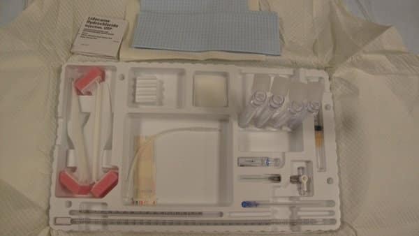
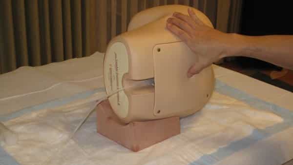
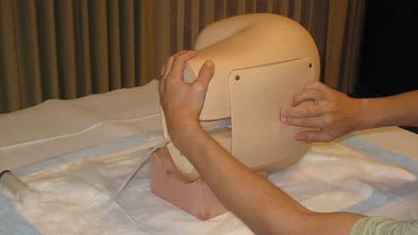
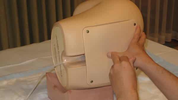
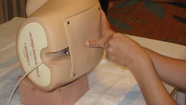
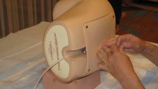
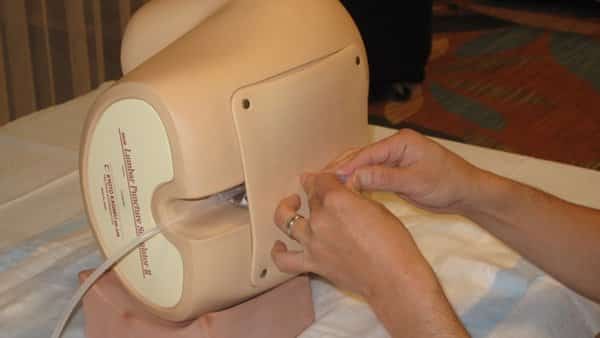
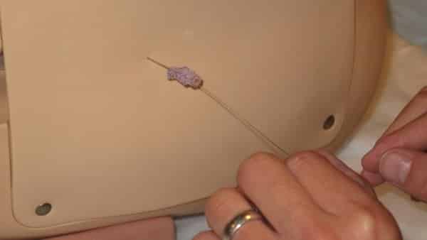
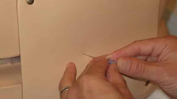
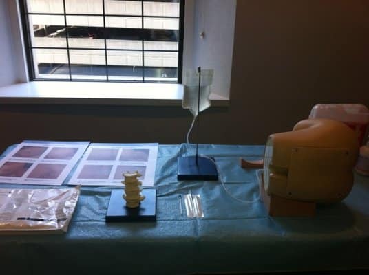
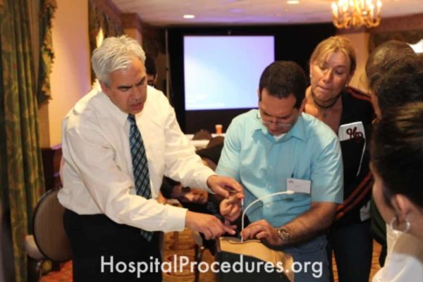
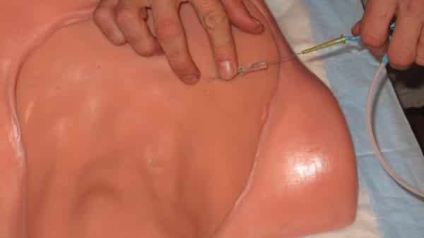
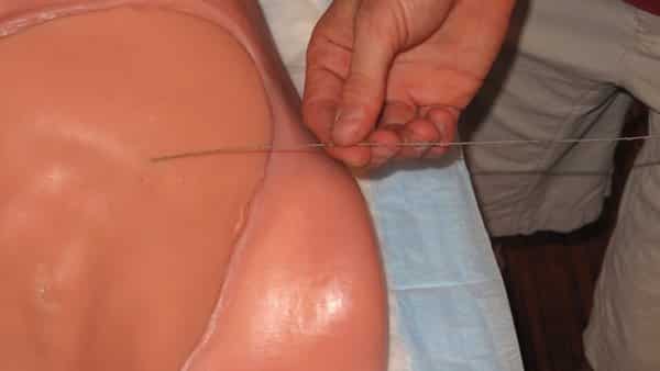
Lumbar Puncture Training
The image depicts a Lumbar Puncture (LP) Course, focusing on educating healthcare professionals in the technique of performing lumbar punctures (spinal taps). This procedure is essential for obtaining cerebrospinal fluid (CSF) for analysis and measuring opening pressure. The course equips medical practitioners with the skills necessary to accurately and safely perform lumbar punctures, addressing critical diagnostic and patient care needs.
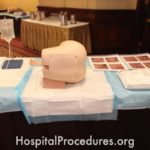
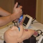
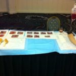
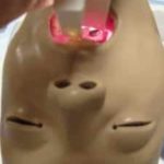
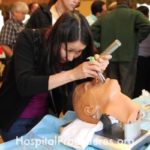
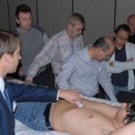
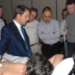
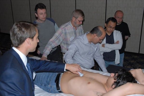
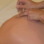
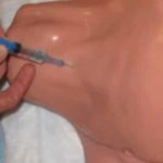
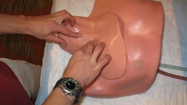
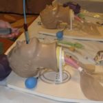
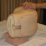
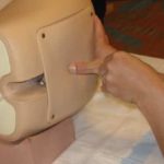
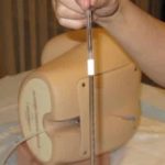
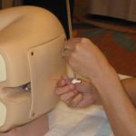
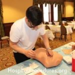
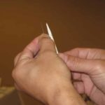
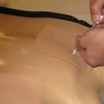
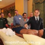
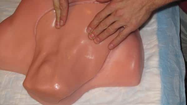
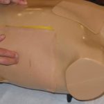
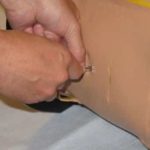
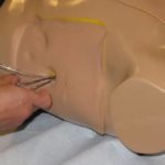
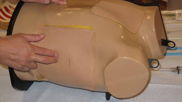
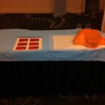
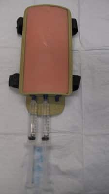
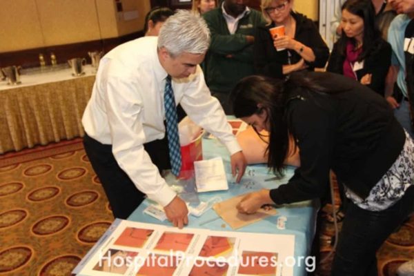
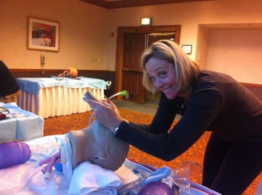
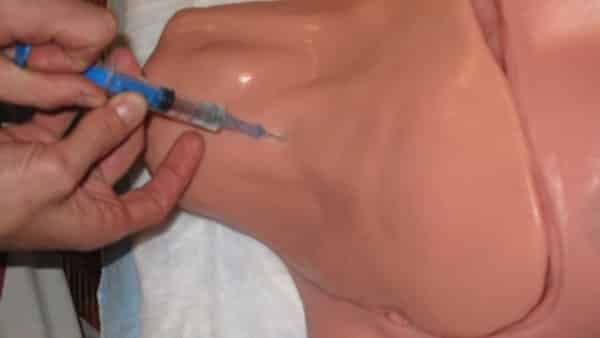
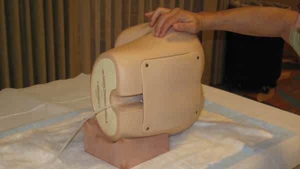
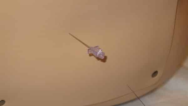
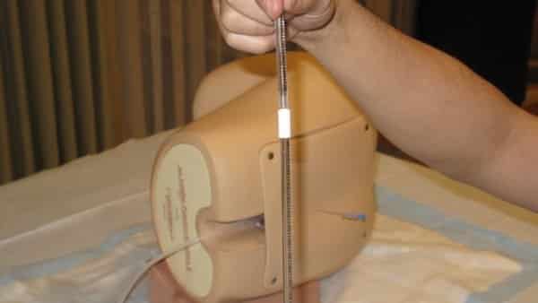
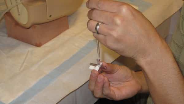
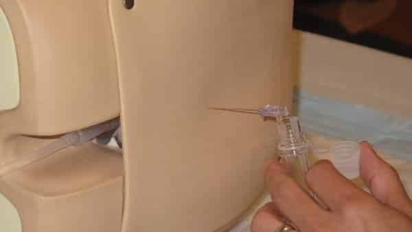
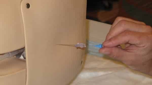
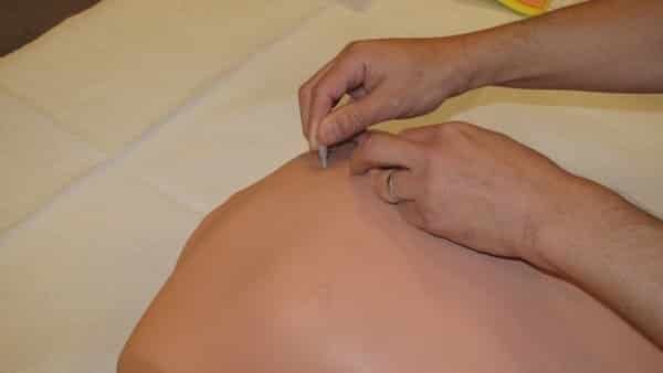
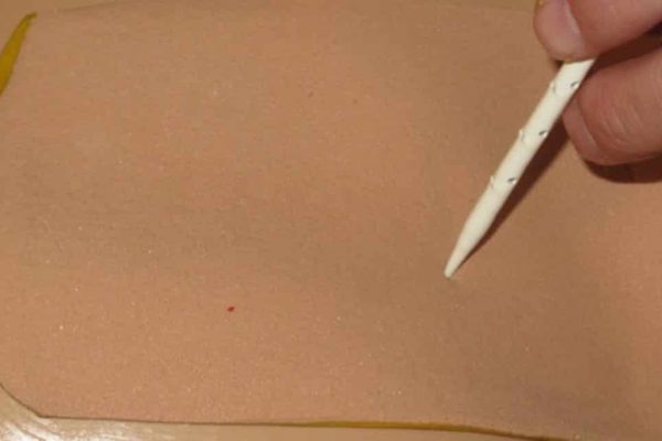
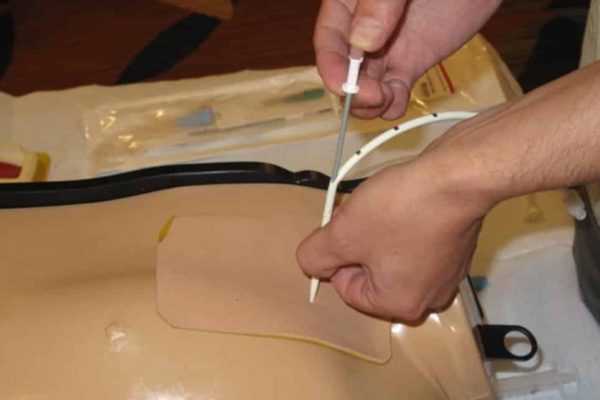
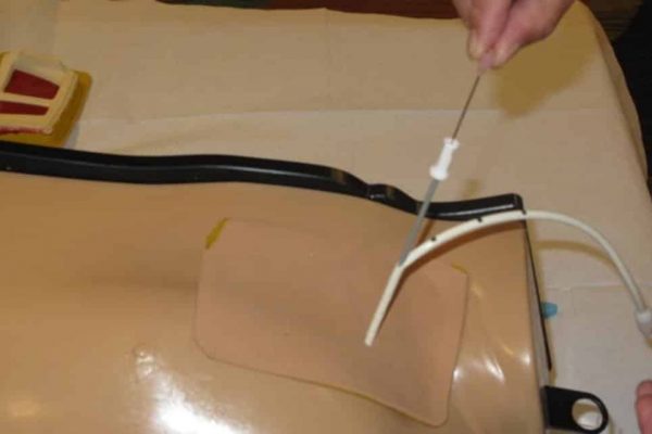
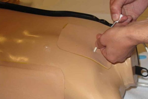
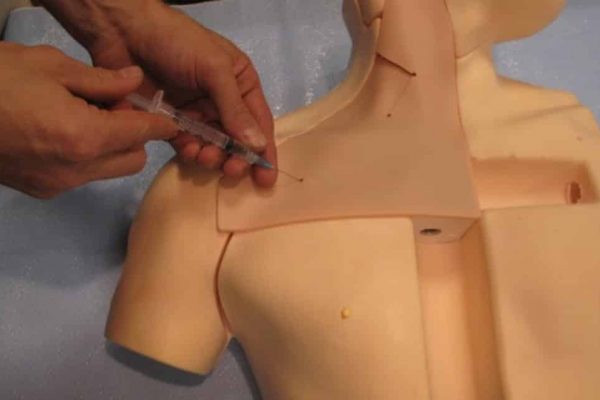
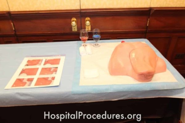
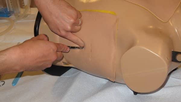
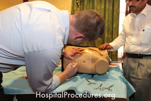
Needle Decompression Course
The image showcases a Needle Decompression Course designed to educate healthcare professionals in the technique of needle decompression. This critical procedure is used to treat tension pneumothorax. The course utilizes an advanced simulator torso to provide comprehensive training in needle decompression at both the mid-clavicular line and the mid-axillary line. The image emphasizes the importance of specialized education for medical practitioners to effectively address tension pneumothorax cases, enhancing patient care and safety.
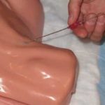
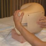
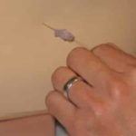
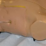
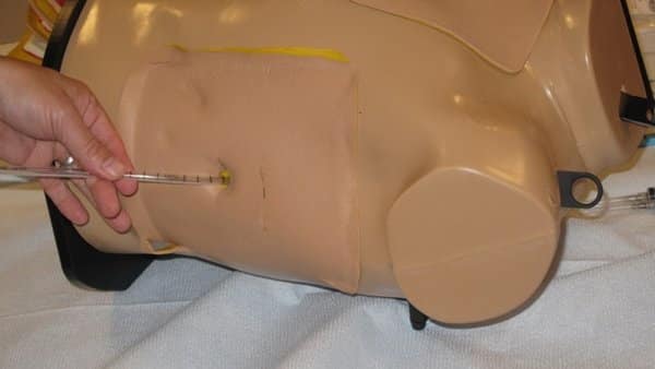
Needle Decompression for Tension Pneumothorax
The image depicts a Needle Decompression procedure being performed to treat a tension pneumothorax. This life-saving intervention is essential in such cases. The description mentions the use of an advanced simulator torso to teach the technique of needle decompression as part of a specialized course. The course covers needle decompression in both the mid-clavicular line and the mid-axillary line approaches. The image underscores the critical nature of needle decompression in managing tension pneumothorax and highlights the value of hands-on training for medical professionals.
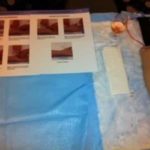
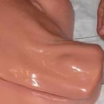
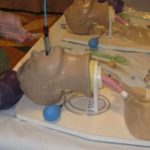
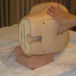
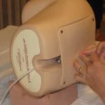
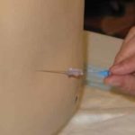
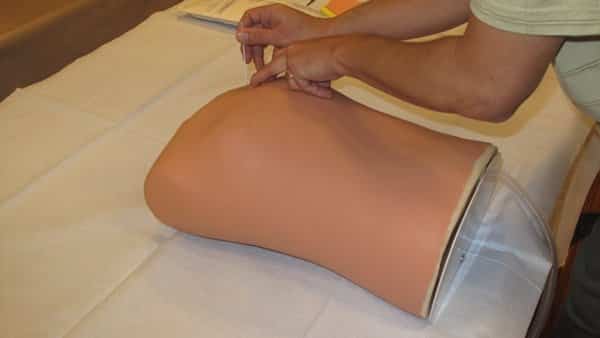
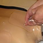
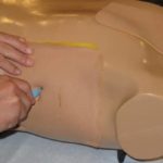
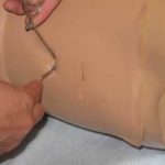
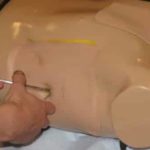
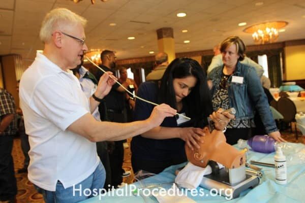
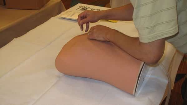
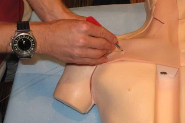
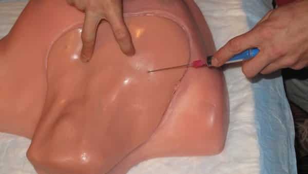
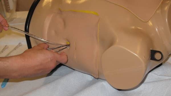
Needle Decompression Landmarks
The image highlights Needle Decompression Landmarks, crucial reference points used for the technique of needle decompression. This procedure is essential for treating tension pneumothorax. The course covers needle decompression in both the mid-clavicular line and the mid-axillary line approaches. The image underscores the significance of accurate landmark identification in ensuring successful and safe needle decompression procedures.
Needle Decompression Course
The image depicts a Needle Decompression Course, dedicated to educating healthcare professionals in the technique of needle decompression. This critical procedure is employed for treating tension pneumothorax. The course employs an advanced simulator torso to offer comprehensive training in needle decompression at both the mid-clavicular line and the mid-axillary line. The image underscores the importance of specialized education to ensure medical practitioners are adept at effectively addressing tension pneumothorax cases, promoting patient safety and optimal outcomes.
Needle Decompression Training
The image represents a Needle Decompression Training session, designed to educate healthcare professionals in the technique of needle decompression. This life-saving procedure is essential for addressing tension pneumothorax. The training utilizes an advanced simulator torso to provide hands-on instruction in performing needle decompression at both the mid-clavicular line and the mid-axillary line. The image underscores the importance of specialized training to ensure medical professionals are proficient in effectively treating tension pneumothorax cases.
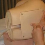
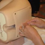
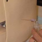
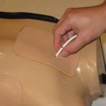
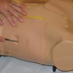
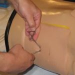
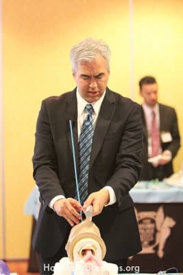
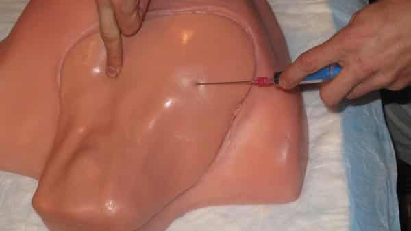
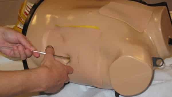
Needle Decompression Technique
The image demonstrates the Needle Decompression Technique, a critical intervention used for treating tension pneumothorax. The accompanying text mentions the utilization of an advanced simulator torso to teach this technique as part of the Needle Decompression Course. The course covers needle decompression in both the mid-clavicular line and the mid-axillary line approaches. The image emphasizes the importance of mastering this skill to effectively address tension pneumothorax cases and underscores the value of specialized training for medical professionals.
Needle Decompression Skills Lab
The image illustrates a Needle Decompression Skills Lab, dedicated to imparting the technique of needle decompression to healthcare professionals. This critical procedure is employed for treating tension pneumothorax. The lab employs an advanced simulator torso to offer specialized training in performing needle decompression at both the mid-clavicular line and the mid-axillary line. The image highlights the importance of a dedicated lab environment for hands-on practice and skill development, enabling medical practitioners to confidently address tension pneumothorax cases.


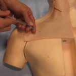
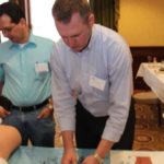
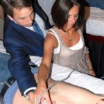
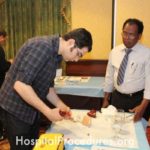
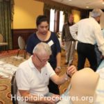
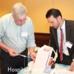
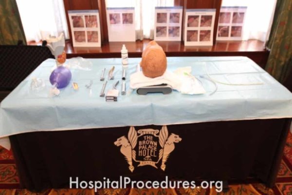
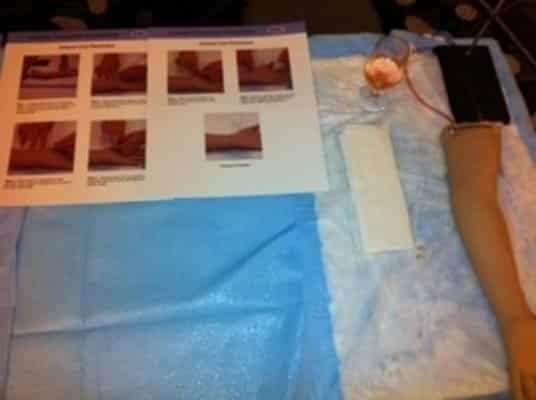
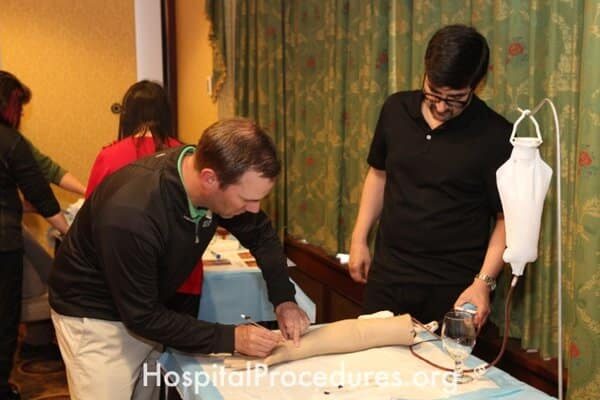
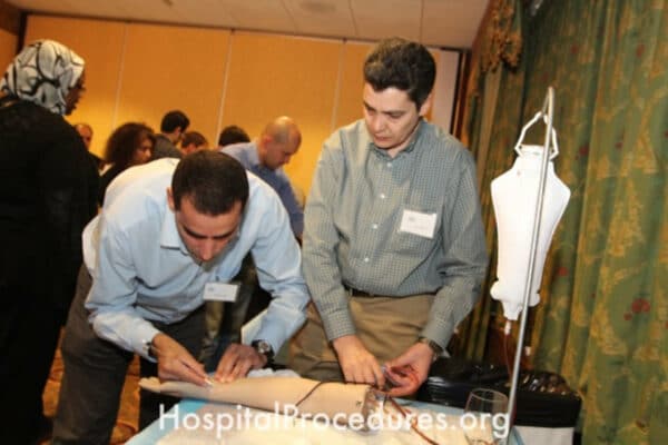
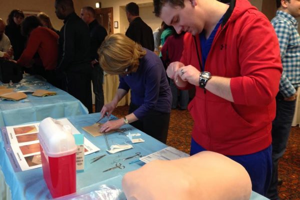
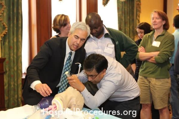
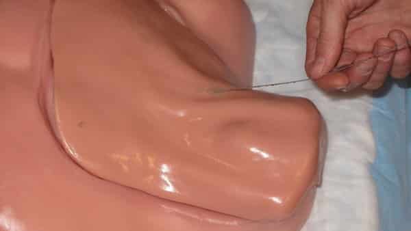
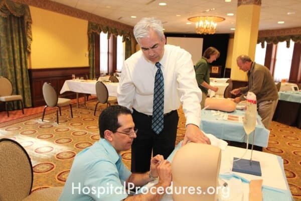
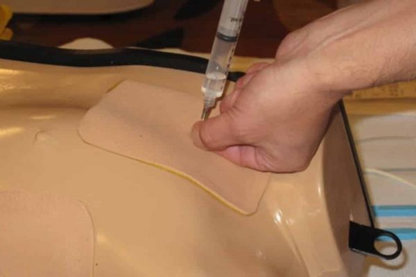
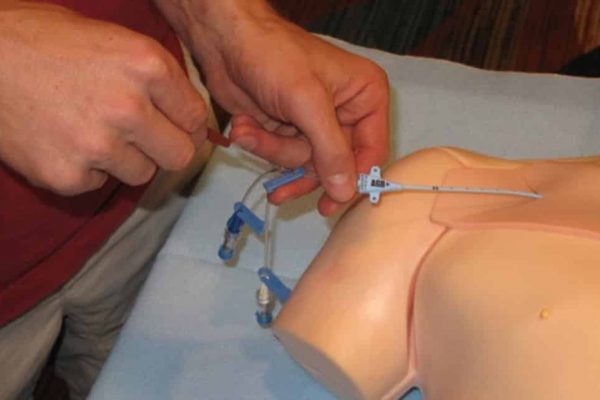
Training for Subclavian Central Venous Catheter Placement
The image captures a training session for subclavian central venous catheter placement. This training is essential for medical professionals to learn the procedure, which is indicated for patients requiring hemodynamic monitoring, rapid volume resuscitation, vasopressor administration, central parenteral nutrition, vesicant infusions, and those with challenging venous access. The procedure enables precise placement of the catheter to facilitate critical medical interventions
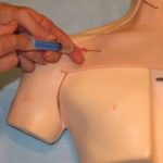
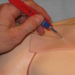
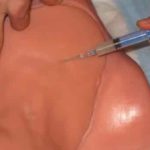
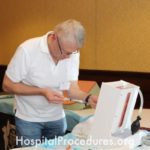
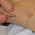
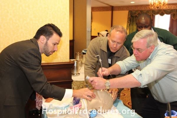
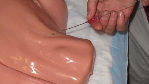
Subclavian Line Insertion Procedure
The image illustrates the process of subclavian line insertion, a medical procedure recommended for patients requiring hemodynamic monitoring, swift volume resuscitation, therapies like vasopressors, central parenteral nutrition, and vesicant infusions. This technique is particularly valuable for patients with challenging venous access. The image captures the essential steps and equipment involved in ensuring successful central line placement.
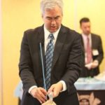
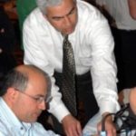
Subclavian Line Continuing Medical Education (CME)
The image features a Continuing Medical Education (CME) session focused on subclavian line placement. This educational opportunity addresses the importance of central line placement for patients requiring hemodynamic monitoring, rapid volume resuscitation, administration of therapies like vasopressors, central parenteral nutrition, and vesicant infusions. It is particularly relevant for patients facing challenging venous access situations, emphasizing skill development and patient safety.

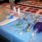
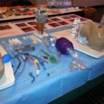
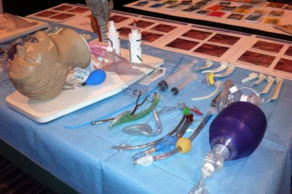
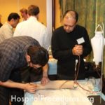
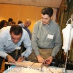
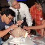
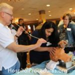
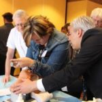
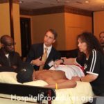
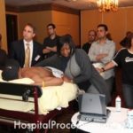
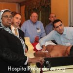
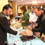
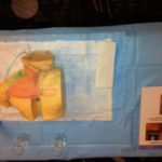
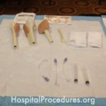
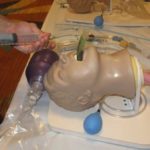
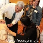
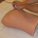
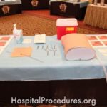
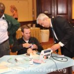
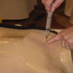
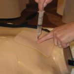
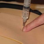
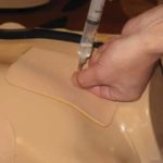
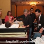
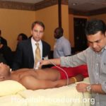
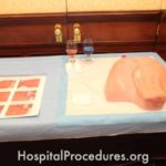
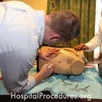
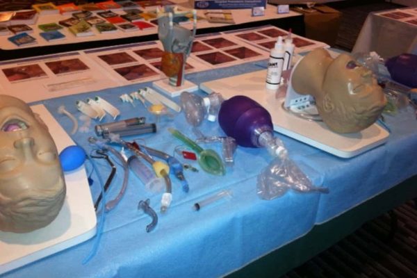
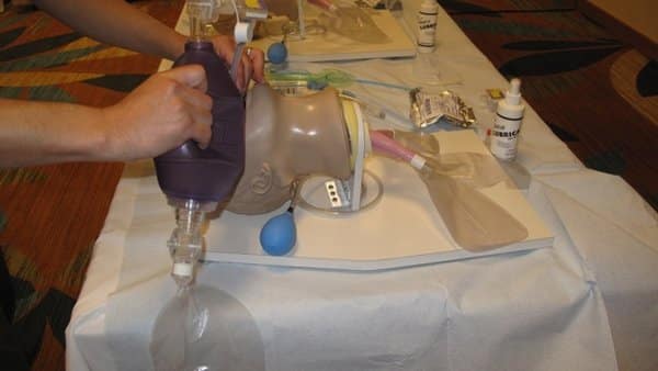
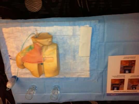
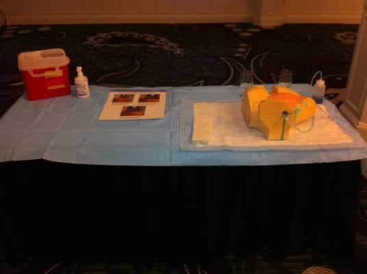
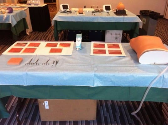
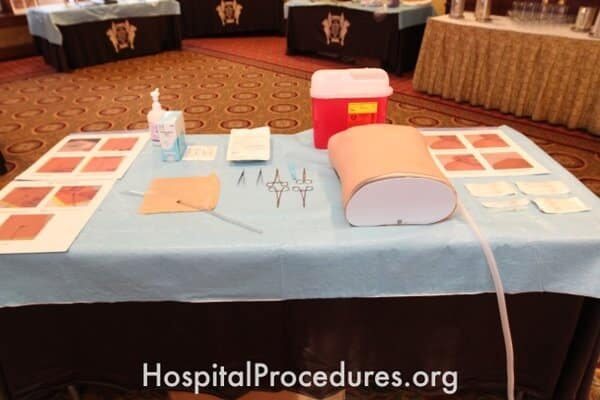
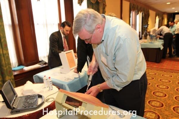
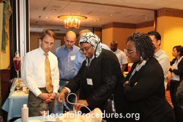
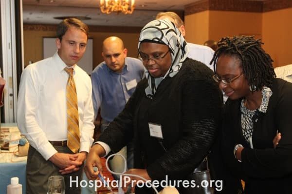
Central Line Placement and Thoracentesis Procedures
The image portrays both a central line placement procedure and a thoracentesis procedure. Central line placement involves the insertion of a central venous catheter for various medical purposes. Thoracentesis, on the other hand, is a procedure used to aspirate pleural fluid from the pleural space. It can serve as a diagnostic method to determine the origin of pleural effusions or to assess for infected fluid. Alternatively, it can be therapeutic, aimed at draining symptomatic pleural effusions. The image captures the critical steps of both procedures, highlighting their importance in comprehensive patient care.
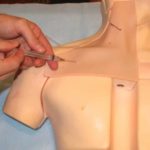
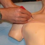
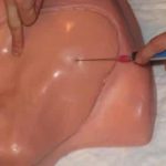
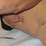
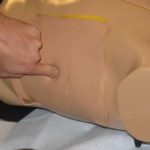
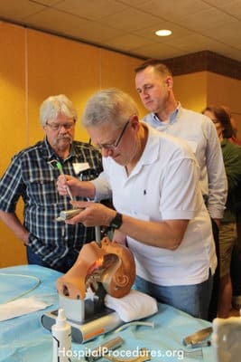
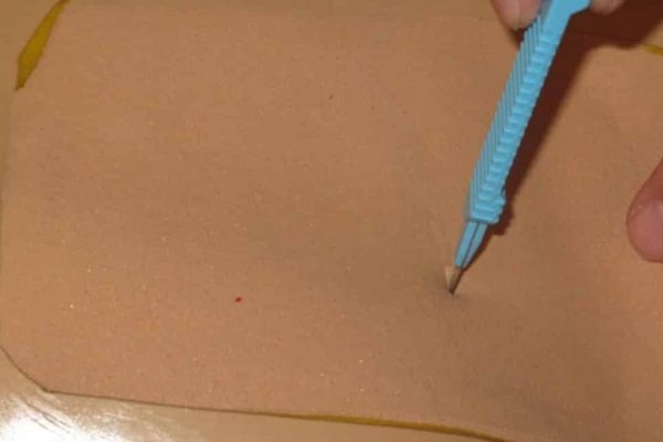
Subclavian Line Landmark Identification
The image demonstrates the identification of anatomical landmarks for subclavian line placement. This procedure is essential for patients requiring hemodynamic monitoring, rapid volume resuscitation, administration of therapies like vasopressors, central parenteral nutrition, and vesicant infusions. Landmark identification is particularly critical for patients with challenging venous access, ensuring accurate and safe central line insertion.
Subclavian Line Insertion Procedure
The image illustrates the process of subclavian line insertion, a medical procedure indicated for patients requiring hemodynamic monitoring, rapid volume resuscitation, administration of therapies like vasopressors, central parenteral nutrition, and vesicant infusions. This technique is particularly valuable for patients with challenging venous access. The image captures the essential steps and equipment involved in ensuring successful central line placement.
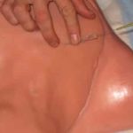
Subclavian Line Placement for Central Venous Access
The image depicts the process of subclavian line placement, a medical procedure indicated for patients requiring hemodynamic monitoring, rapid volume resuscitation, vasopressor administration, central parenteral nutrition, and challenging venous access. This technique allows for central venous access, facilitating the delivery of essential medical treatments.
Subclavian Line Placement Course
The image showcases a comprehensive course on subclavian line placement. This course addresses the necessity of central line placement for patients requiring hemodynamic monitoring, swift volume resuscitation, administration of therapies like vasopressors, central parenteral nutrition, and vesicant infusions. It holds particular significance for patients with challenging venous access, providing participants with in-depth knowledge and hands-on training to proficiently perform this critical medical procedure.
Subclavian Line CME Seminar
The image portrays a Continuing Medical Education (CME) seminar focusing on subclavian line placement. This educational event emphasizes the importance of central line placement for patients necessitating hemodynamic monitoring, rapid volume resuscitation, administration of therapies like vasopressors, central parenteral nutrition, and vesicant infusions. It is especially relevant for patients with challenging venous access situations, providing healthcare professionals with valuable insights and expertise in performing this essential procedure.
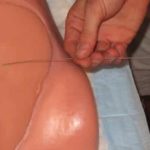
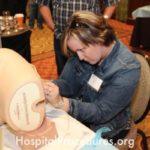
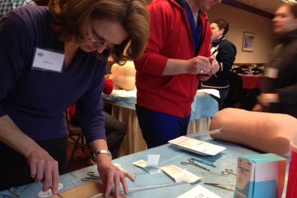
Subclavian Line Training Session
The image showcases a training session focused on subclavian line placement. This training addresses the significance of central line placement for patients necessitating hemodynamic monitoring, rapid volume resuscitation, administration of therapies like vasopressors, central parenteral nutrition, and vesicant infusions. It is particularly beneficial for patients with challenging venous access, offering healthcare professionals the skills needed to perform this procedure effectively and ensure patient well-being.

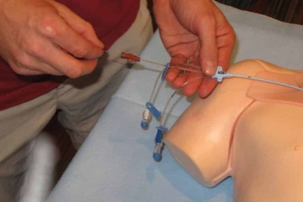
Training for Subclavian Central Venous Catheter Placement
The image illustrates a training session for subclavian central venous catheter placement. This procedure is essential for patients requiring hemodynamic monitoring, rapid volume resuscitation, administering therapies like vasopressors, central parenteral nutrition, and vesicant infusions. It is particularly crucial for patients with challenging venous access, allowing medical professionals to gain the necessary skills for precise catheter placement.
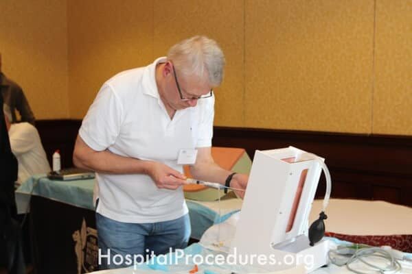
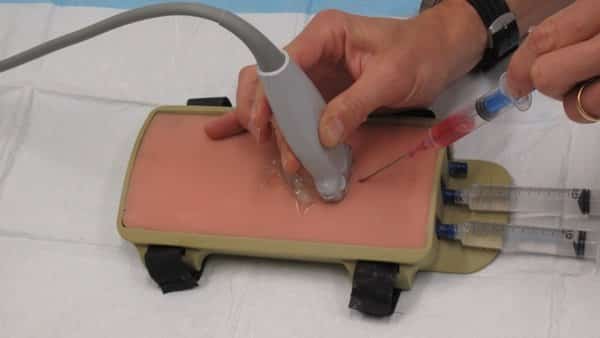
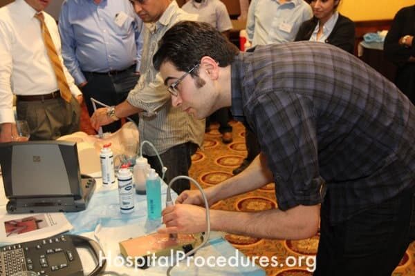
Ultrasound-Guided Subclavian Line Procedure
The image depicts the skilled execution of an ultrasound-guided subclavian line insertion. This advanced technique is crucial for patients requiring hemodynamic monitoring, swift volume resuscitation, vasopressor administration, central parenteral nutrition, and vesicant infusions. Especially beneficial for patients with challenging venous access, ultrasound guidance enhances accuracy and safety during the central line placement process.


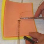
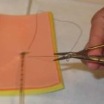
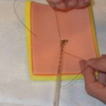
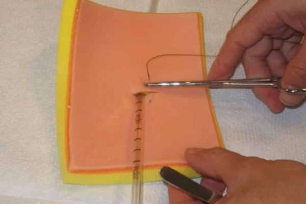
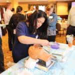
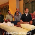
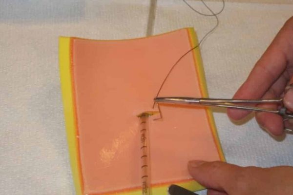
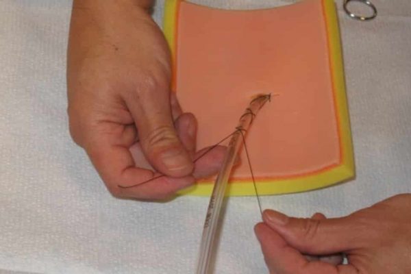
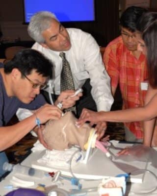
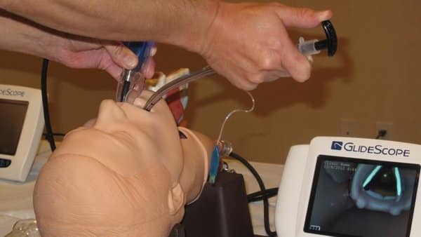
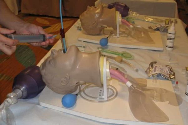
Tube Thoracostomy Course: Mastering Chest Tube Placement
The image depicts a dedicated Tube Thoracostomy Course, focusing on educating healthcare professionals in the art of chest tube placement. This technique, known as tube thoracostomy, is essential for managing conditions like pneumothorax, hemothorax, empyema, complicated parapneumonic effusions, and supporting pleurodesis procedures. The course equips medical practitioners with the knowledge and skills necessary to skillfully insert chest tubes, addressing critical thoracic conditions and providing comprehensive patient care.
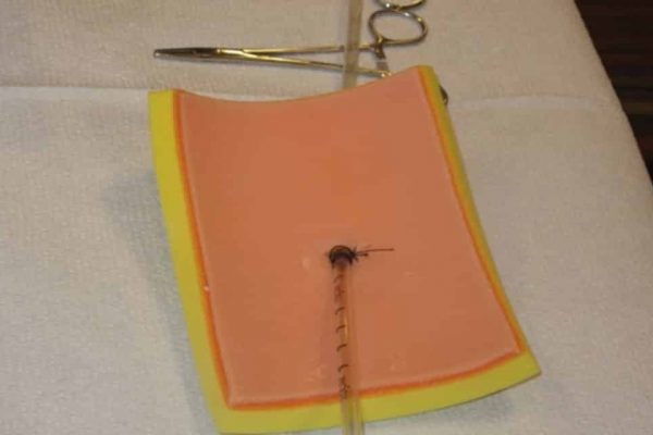
Tube Thoracostomy Continuing Medical Education (CME)
The image represents a Continuing Medical Education (CME) session focused on tube thoracostomy, also known as chest tube placement. This educational event emphasizes the significance of this procedure for treating conditions such as pneumothorax, hemothorax, empyema, complicated parapneumonic effusions, and facilitating pleurodesis. The CME equips healthcare professionals with comprehensive knowledge and skills to perform tube thoracostomy accurately and safely, ensuring optimal patient care and outcomes.
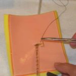
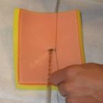
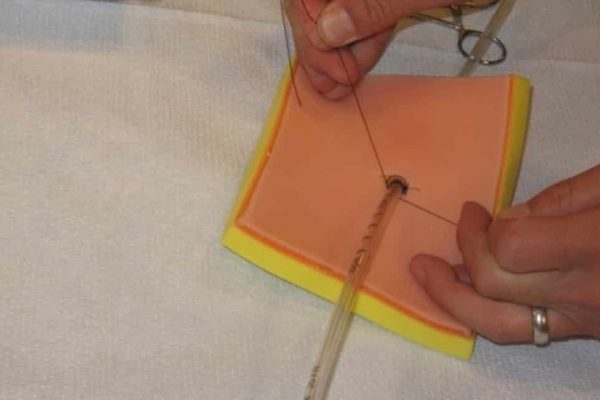
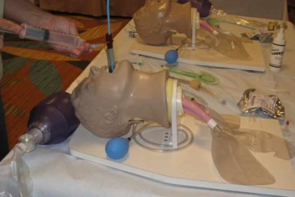
Tube Thoracostomy Insertion Procedure
The image illustrates the process of tube thoracostomy insertion, also known as chest tube placement. This procedure is essential for treating conditions such as pneumothorax, hemothorax, empyema, complicated parapneumonic effusions, and to assist in performing pleurodesis. The image captures the precise steps and equipment involved in safely and accurately inserting a chest tube, highlighting its importance in managing various thoracic conditions and promoting optimal patient recovery.
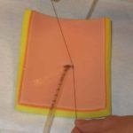
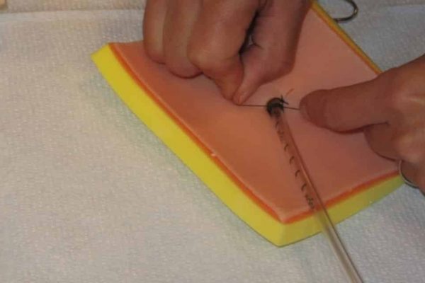
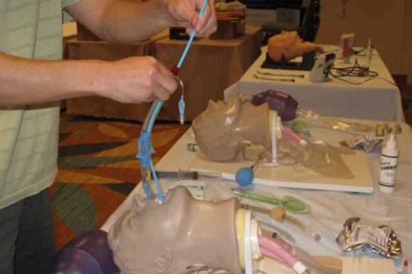
Tube Thoracostomy Insertion Procedure
The image illustrates the process of tube thoracostomy insertion, commonly known as chest tube placement. This procedure is crucial for treating conditions such as pneumothorax, hemothorax, empyema, complicated parapneumonic effusions, and facilitating pleurodesis. The image captures the steps involved in accurately and safely inserting a chest tube, addressing critical patient care needs in various thoracic conditions.
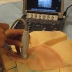
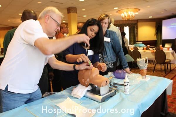
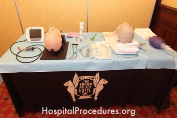
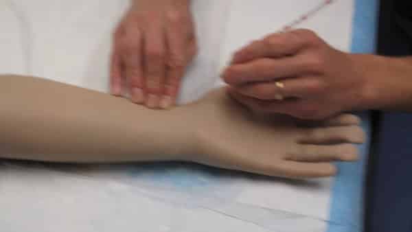
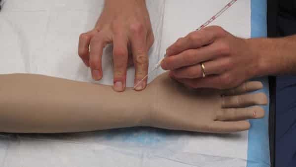
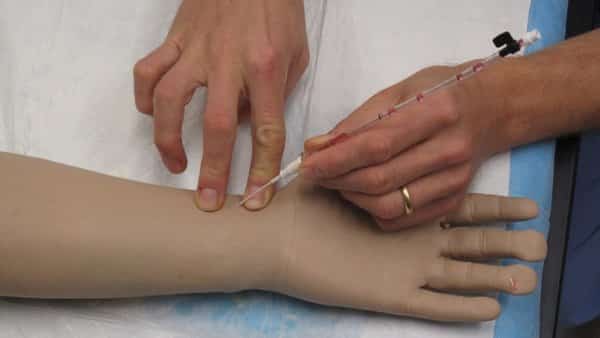
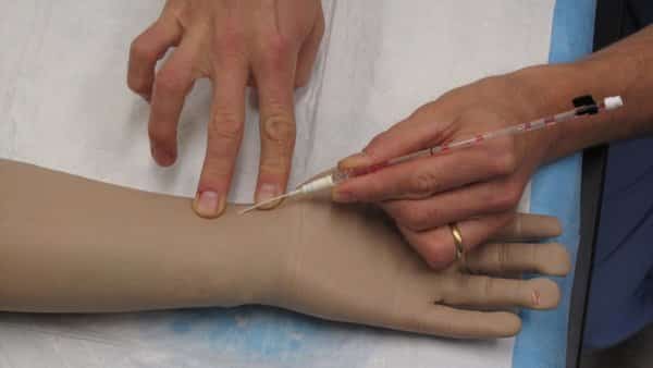
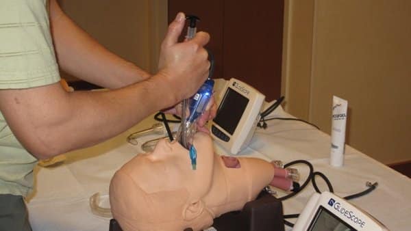
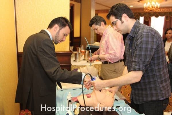
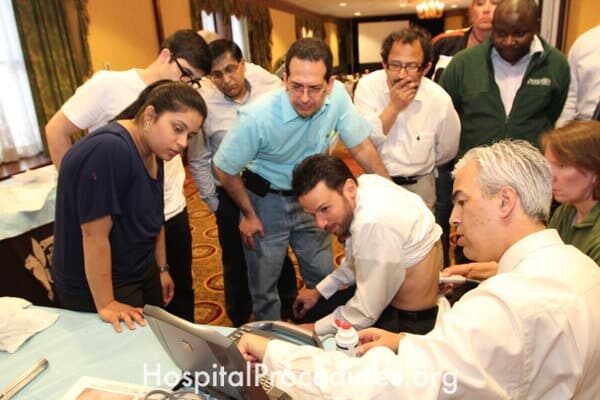
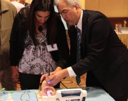
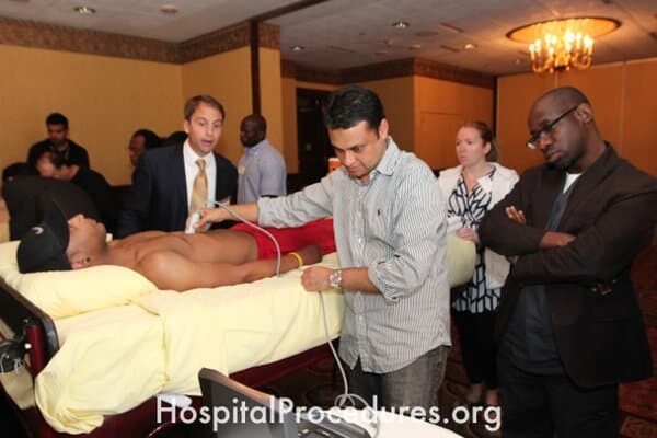
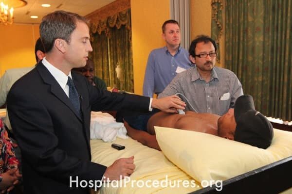
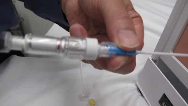
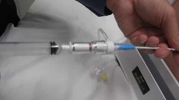
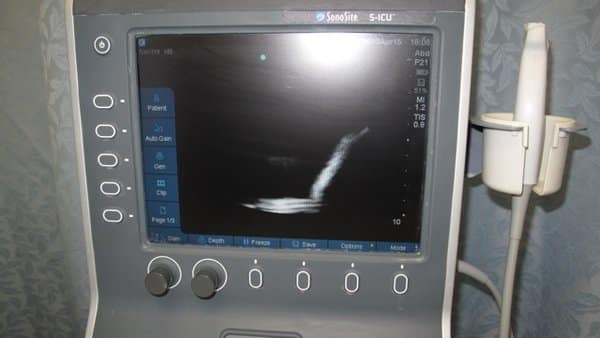
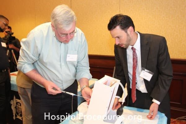
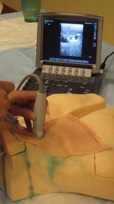
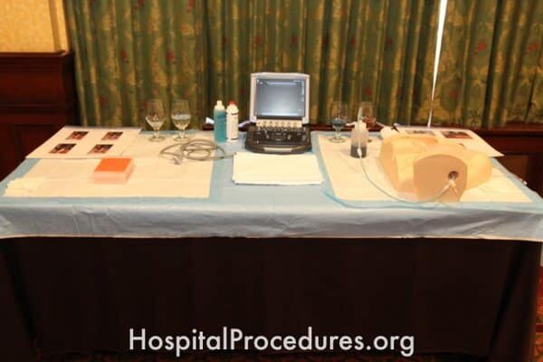
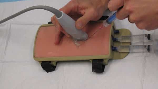
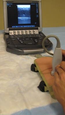
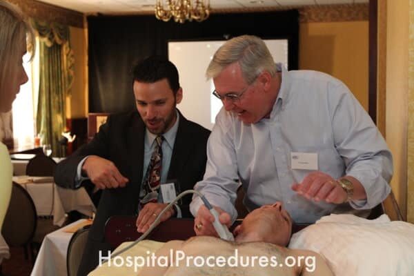
Ultrasound-Guided Central Line Insertion
The image showcases the utilization of ultrasound guidance for the insertion of central lines. This technique is vital for patients requiring hemodynamic monitoring, rapid volume resuscitation, administration of therapies like vasopressors, central parenteral nutrition, and vesicant infusions. The image also represents an Ultrasound-Guided Central Line Course, emphasizing its significance in enhancing skills for healthcare professionals to proficiently perform central line placements, particularly in patients with challenging venous access.

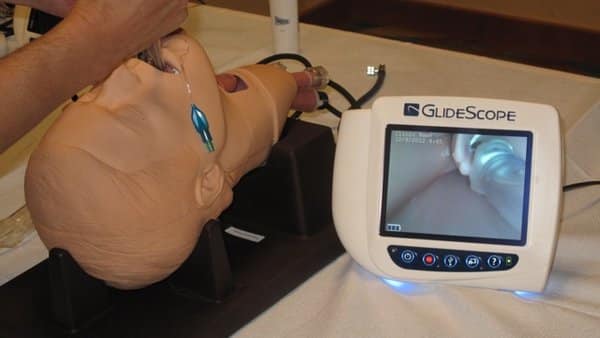
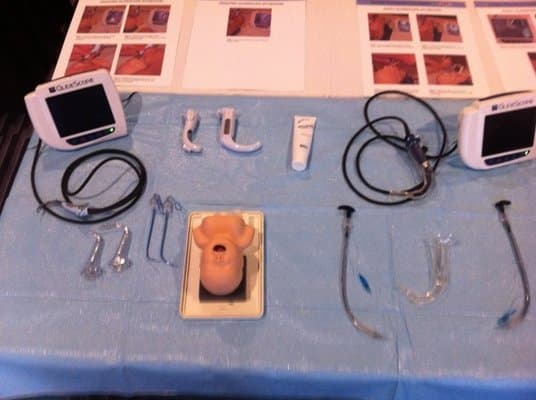
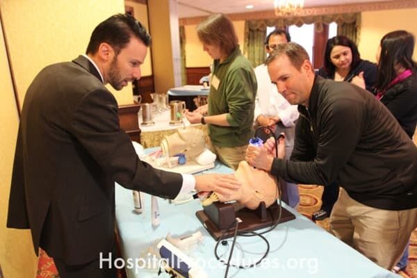
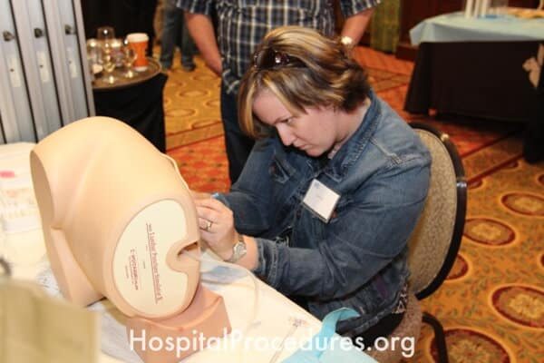
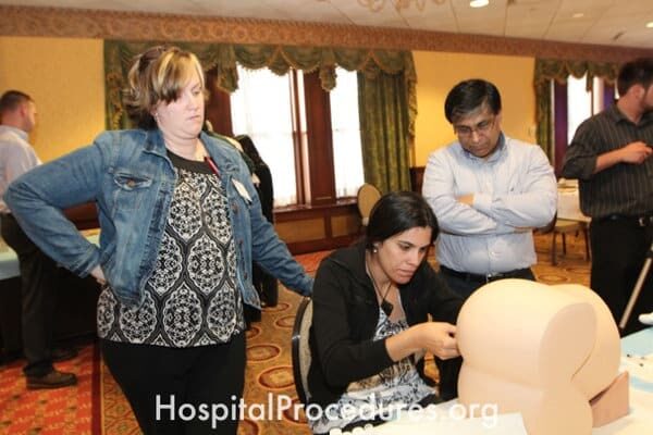
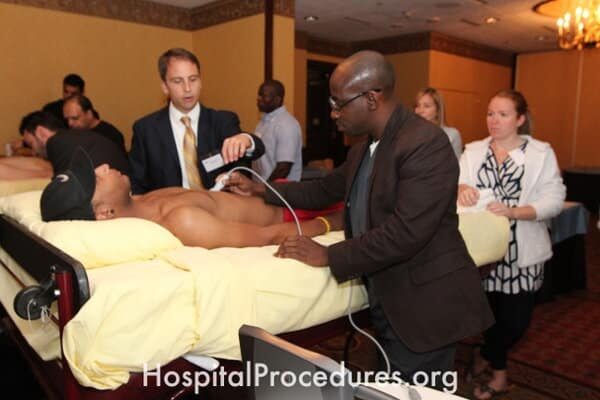
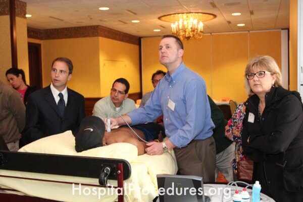
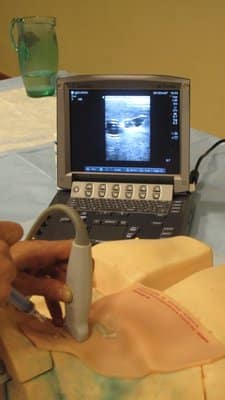
Ultrasound-Guided Central Line Lab
The image features an Ultrasound-Guided Central Line Lab, part of the comprehensive Central Line Course. This educational setting focuses on training healthcare professionals in the use of ultrasound guidance for central line placement. The course addresses the needs of patients requiring hemodynamic monitoring, rapid volume resuscitation, administration of therapies like vasopressors, central parenteral nutrition, and vesicant infusions. It is especially beneficial for patients with challenging venous access, providing hands-on experience to ensure precise and safe central line insertion.
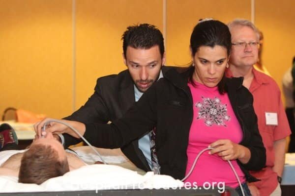
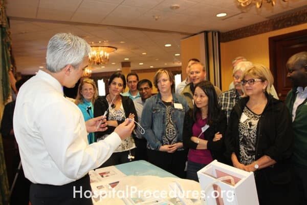
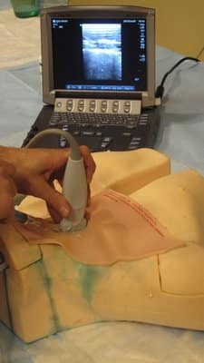
Ultrasound-Guided Central Line Lab Training
The image captures a hands-on training session in the Ultrasound-Guided Central Line Lab Course. This specialized course focuses on teaching the technique of central line placement under ultrasound guidance. The training caters to patients requiring hemodynamic monitoring, rapid volume resuscitation, administration of therapies like vasopressors, central parenteral nutrition, and vesicant infusions. It is particularly designed for patients with challenging venous access, offering healthcare professionals practical skills to ensure precise and safe central line insertion.

Ultrasound-Guided Central Line Lab
The image showcases an Ultrasound-Guided Central Line Course laboratory session. This educational setting focuses on teaching healthcare professionals the technique of using ultrasound guidance for central line placement. The course addresses the critical needs of patients requiring hemodynamic monitoring, swift volume resuscitation, administration of therapies like vasopressors, central parenteral nutrition, and vesicant infusions. It especially caters to patients with challenging venous access, empowering participants with hands-on skills to ensure accurate and safe central line insertion.


